. 2003 May 5;197(9):1107-17.
doi: 10.1084/jem.20021787. Epub 2003 Apr 28.

Collaborative induction of inflammatory responses by dectin-1 and Toll-like receptor 2

Affiliations

Collaborative induction of inflammatory responses by dectin-1 and Toll-like receptor 2

Benjamin N Gantner et al. J Exp Med. .

Abstract

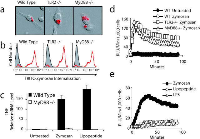
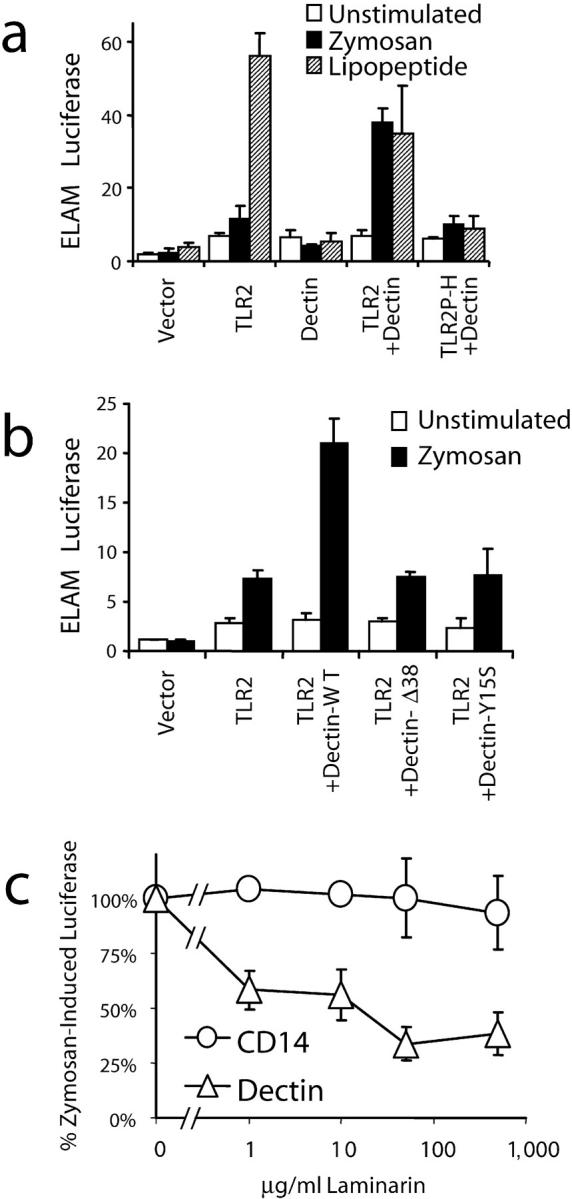
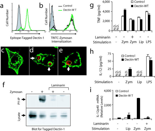
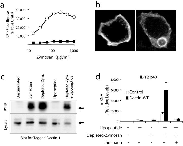
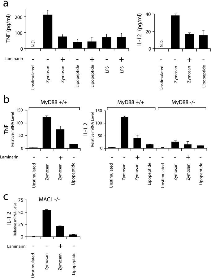
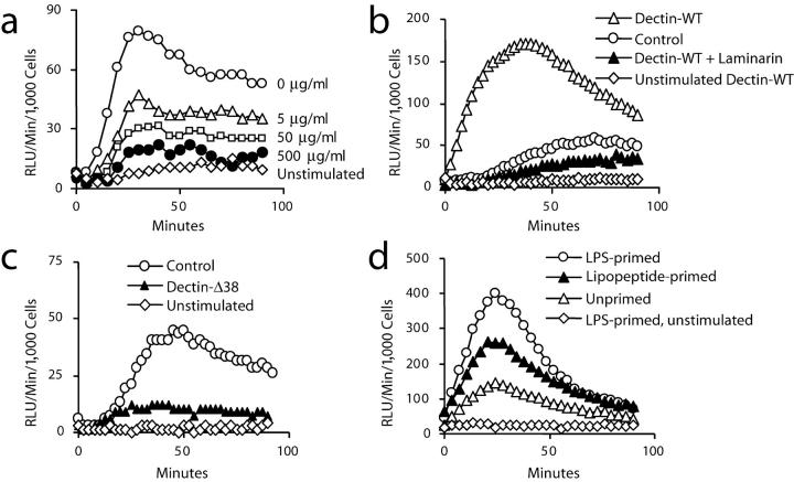
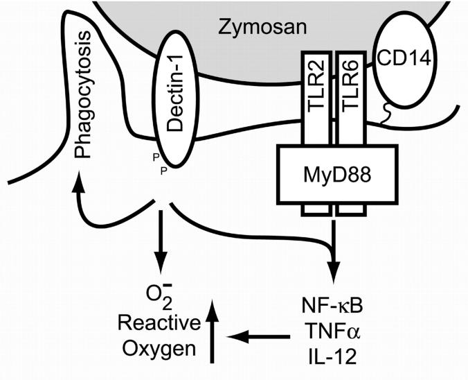
Toll-like receptors (TLRs) mediate recognition of a wide range of microbial products including lipopolysaccharides, lipoproteins, flagellin, and bacterial DNA, and signaling through TLRs leads to the production of inflammatory mediators. In addition to TLRs, many other surface receptors have been proposed to participate in innate immunity and microbial recognition, and signaling through some of these receptors is likely to cooperate with TLR signaling in defining inflammatory responses. In this report we have examined how dectin-1, a lectin family receptor for beta-glucans, collaborates with TLRs in recognizing microbes. Dectin-1, which is expressed at low levels on macrophages and high levels on dendritic cells, contains an immunoreceptor tyrosine-based activation motif-like signaling motif that is tyrosine phosphorylated upon activation. The receptor is recruited to phagosomes containing zymosan particles but not to phagosomes containing immunoglobulin G-opsonized particles. Dectin-1 expression enhances TLR-mediated activation of nuclear factor kappa B by beta-glucan-containing particles, and in macrophages and dendritic cells dectin-1 and TLRs are synergistic in mediating production of cytokines such as interleukin 12 and tumor necrosis factor alpha. Additionally, dectin-1 triggers production of reactive oxygen species, an inflammatory response that is primed by TLR activation. The data demonstrate that collaborative recognition of distinct microbial components by different classes of innate immune receptors is crucial in orchestrating inflammatory responses.


Figures

Figure 1.
Figure 1.
Zymosan engages receptors on macrophages other than TLRs. (a and b) Bone marrow–derived macrophages from wild-type, TLR2−/−, or MyD88−/− mice were incubated with tetramethylrhodamine isothiocyanate–labeled zymosan for 15 min. Phagocytosis was visualized by immunofluorescence microscopy (a) and quantified by flow cytometry (b). (c) Bone marrow macrophages from MyD88−/− or wild-type mice were stimulated with 100 μg/ml zymosan or 100 ng/ml PAM3CSK4 lipopeptide for 4 h and TNF-α mRNA production was measured by quantitative real-time PCR. (d and e) Production of ROS was assayed by luminol-enhanced chemiluminescence in peritoneal macrophages from wild-type, TLR2−/−, or MyD88−/− mice (d) stimulated with 200 μg/ml zymosan or in RAW 264.7 macrophages (e) stimulated with 100 μg/ml zymosan, 100 ng/ml LPS, or 100 ng/ml PAM3CSK4 lipopeptide.
Figure 2.
Figure 2.
Dectin-1 and TLR2 collaborate in zymosan recognition by HEK 293 cells. (a and b) Activation of NF-κB (ELAM luciferase) was measured in HEK 293 cells transiently transfected with the indicated expression vectors and stimulated with 100 μg/ml zymosan or 100 ng/ml PAM3CSK4 lipopeptide for 4 h. (c) Activation of NF-κB was measured in HEK 293 cells transfected with expression vectors for TLR2 (together with either CD14 or dectin-1) and stimulated with 100 μg/ml zymosan in the indicated concentrations of laminarin.
Figure 3.
Figure 3.
Zymosan triggers activation of dectin-1 and enhanced inflammatory responses in mouse macrophages. (a) V5 epitope–tagged dectin-1 expression was measured by flow cytometry in RAW cells stably overexpressing wild-type dectin-1 (green line) and in control RAW cells (shaded). (b) Control cells (black line) and cells overexpressing dectin-1 (green line) were incubated with tetramethylrhodamine isothiocyanate zymosan for 1 h and phagocytosis was measured by flow cytometry and compared with unfed control cells (shaded). (c–e) The cellular distribution of epitope-tagged dectin-1 (green) was examined by immunofluorescence microscopy in resting cells (c), cells fed zymosan (red) for 5 min (d), or in cells fed IgG-opsonized sheep red blood cells (red) for 5 min (e). (f) Cells expressing V5-tagged dectin-1 were fed 100 μg/ml zymosan for 15 min in the presence or absence of 50 μg/ml laminarin, total cell lysates were prepared, and the protein was detected in all lysates by immunoblot using an antibody for the tag (bottom). Tyrosine-phosphorylated proteins were immunoprecipitated from the lysates and recovery of dectin-1 was analyzed by immunoblot (top). (g and h) Induction of inflammatory responses was measured by ELISA in control RAW cells and cells overexpressing dectin-1 stimulated with 100 μg/ml zymosan (zym), 100 ng/ml PAM3CSK4 lipopeptide (Lip), or 100 ng/ml LPS for 4 (g, TNF-α) or 24 h (h, IL-12 p40) in the presence or absence of 500 μg/ml laminarin as indicated. N.D., none detected. (i) Induction of IL-12 p40 mRNA was measured by quantitative real-time PCR in cells stimulated for 4 h as in g.
Figure 4.
Figure 4.
Zymosan depleted of its TLR stimulatory activity synergizes with a TLR2 agonist through activation of dectin-1. (a) RAW cells stably transfected with an NF-κB luciferase reporter (ELAM luciferase) were stimulated for 4 h with the indicated concentrations of zymosan (○) or depleted zymosan (▪), and luciferase activity was measured. (b) HEK 293 cells were transiently transfected with dectin-1 fused to green fluorescent protein at the receptor's intracellular amino terminus and dectin-1 localization was visualized before (left) and after (right) exposure to depleted zymosan. (c) Cells expressing V5-tagged dectin-1 were stimulated with 100 μg/ml zymosan, 100 μg/ml depleted zymosan, or PAM3CSK4 lipopeptide as indicated for 15 min in the presence or absence of 50 μg/ml laminarin. Total cell lysates were prepared and the protein was detected by immunoblot using an antibody for the tag (bottom). Tyrosine-phosphorylated proteins were immunoprecipitated from the lysates and recovery of dectin-1 was analyzed by immunoblot (top). (d) IL-12 p40 mRNA production was measured by quantitative real-time PCR in control RAW cells and cells overexpressing dectin-1 stimulated for 4 h with 100 ng/ml PAM3CSK4 lipopeptide or 100 μg/ml depleted zymosan in the presence or absence of 500 μg/ml laminarin.
Figure 5.
Figure 5.
TLRs and dectin-1 collaborate in primary bone marrow dendritic cells. (a) Induction of cytokines in mouse BMDCs was measured by ELISA. The cells were stimulated for 4 h with 100 μg/ml zymosan or 100 ng/ml PAM3CSK4 lipopeptide in the presence or absence of 500 μg/ml laminarin. N.D., none detected. (b) Induction of cytokines was measured by quantitative real-time PCR in primary dendritic cells from MyD88+/+ and MyD88−/− mice stimulated for 4 h as in a. (c) Induction of IL-12 p40 was measured by quantitative real-time PCR in dendritic cells from Mac-1(CD11b)−/− mice stimulated as in b.
Figure 6.
Figure 6.
Dectin-1 stimulates production of ROS in macrophages. (a) Production of ROS was assayed by luminol-enhanced chemiluminescence in RAW cells in the presence of 100 μg/ml zymosan together with the indicated concentrations of laminarin. (b) Zymosan-induced production of ROS in RAW cells was compared with RAW cells overexpressing wild-type dectin-1 in the presence or absence of 500 μg/ml laminarin. (c) Zymosan-induced production of ROS in RAW cells was compared with RAW cells overexpressing a signaling-deficient (Δ38) form of dectin-1. (d) Zymosan-induced production of ROS was measured in RAW cells overexpressing wild-type dectin-1 with or without 4 h prestimulation with 100 ng/ml LPS or 100 ng/ml PAM3CSK4 lipopeptide.
Figure 7.
Figure 7.
Model of dectin-1/TLR collaboration. Zymosan particles are recognized simultaneously by dectin-1, TLRs, and CD14. Together these receptors facilitate inflammatory responses to the particle. Dectin-1 directly triggers phagocytosis and stimulates the production of ROS, thus contributing to microbial killing. TLRs induce signaling through NF-κB that leads to the production of inflammatory cytokines and these responses are enhanced by dectin-1. Similarly, TLR signaling enhances responses such as the production of ROS that are triggered by dectin-1.

References

    1. Underhill, D.M., and A. Ozinsky. 2002. Phagocytosis of microbes: complexity in action. Annu. Rev. Immunol. 20:825–852. - PubMed
    1. Henson, P.M., D.L. Bratton, and V.A. Fadok. 2001. Apoptotic cell removal. Curr. Biol. 11:R795–R805. - PubMed
    1. Underhill, D.M., and A. Ozinsky. 2002. Toll-like receptors: key mediators of microbe detection. Curr. Opin. Immunol. 14:103–110. - PubMed
    1. Aderem, A., and R.J. Ulevitch. 2000. Toll-like receptors in the induction of the innate immune response. Nature. 406:782–787. - PubMed
    1. Akira, S., K. Takeda, and T. Kaisho. 2001. Toll-like receptors: critical proteins linking innate and acquired immunity. Nat. Immunol. 2:675–680. - PubMed

Publication types