. 2003 May 13;100(10):5938-43.
doi: 10.1073/pnas.1031791100. Epub 2003 Apr 28.

Functional conservation of light, oxygen, or voltage domains in light sensing

Affiliations

Functional conservation of light, oxygen, or voltage domains in light sensing

Ping Cheng et al. Proc Natl Acad Sci U S A. .

Abstract

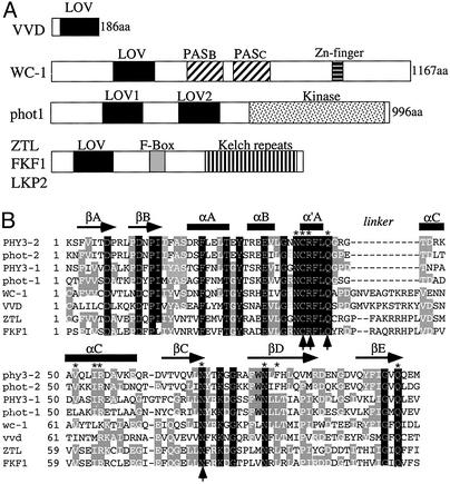
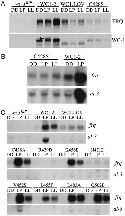
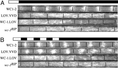
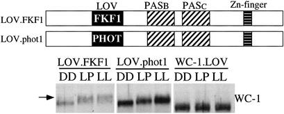
In Neurospora, the flavin adenine dinucleotide-containing protein WHITE COLLAR-1 is the blue-light photoreceptor for the circadian clock and other light responses. The putative chromophore-binding domain of WC-1, its light, oxygen, or voltage (LOV) domain, is similar to the LOV domains found in the plant phototropins, the Neurospora VIVID (VVD) protein, and the Arabidopsis FKF1 and its related proteins. Studies of the plant phototropins have identified 11 flavin-contacting residues that are also conserved in the LOV domains of WC-1, VVD, and FKF1. In this study, by mutating the putative WC-1 flavin-binding sites, we show that these sites are important for the light function of the protein, suggesting that the WC-1 LOV domain adapts a structure similar to that of the phototropin LOV domains. By creating a Neurospora strain in which the LOV domain of WC-1 is swapped with that of VVD, we show that the LOV domain of VVD partially replaces the function of the WC-1 LOV domain, suggesting that VVD is a wc-dependent photoreceptor in Neurospora. Furthermore, we show that the Neurosporastrains containing a chimeric WC-1 protein with the LOV domain from FKF1 or phot1 can also sense light, suggesting that FKF1 and its related proteins are light sensors in Arabidopsis. Taken together, our data suggest that these LOV domains are structurally similar protein modules involved in blue-light sensing.


Figures

Figure 1
Figure 1
(A) Schematic depictions of the domain structures of WC-1, VVD, phot1, and the ZTL family member proteins. (B) Amino acid sequence alignment of LOV domains from phototropins, WC-1, VVD, ZTL, and FKF1. This alignment was modified from ref. and uses the same nomenclature for structural elements. Both LOV domains of the Adiantum capillus-veneris PHY3 and Arabidopsis thaliana phot1 were used in the alignment. Black residues are identical in all LOV domains, and gray residues are identical in most LOV domains. Asterisks mark the 11 FMN-interacting residues found in the LOV2 domain of Adiantum PHY3. Arrows mark the four residues that are essential for the light function of WC-1. For simplicity, the first residue of each LOV domain was numbered as the first residue.
Figure 2
Figure 2
The putative flavin-binding sites of the WC-1 LOV domain are important for its light function. (A and B) Western blot (A) and Northern (B) analyses showing that the C428S mutant is not light-sensitive. WC1-2 is a wc-1RIP strain that carries the wild-type wc-1 construct. In the WC1.LOV strain, the entire WC-1 LOV domain is deleted (15). DD, cultures were harvested after incubation for 24 h in constant darkness (DD24). Light pulse (LP), cultures were exposed to a 15-min light pulse (light intensity, ≈2,000 lux) at DD24. LL, cultures were grown in constant light for >1 day. In A, the arrow indicates the hyperphosphorylated WC-1 of the WC1-2 strain after a light pulse. (C) Northern analyses showing the light induction of frq and al-3 in various wc-1 mutants that contain a single mutation of the putative flavin-binding sites.
Figure 3
Figure 3
The LOV domain of VVD can partially replace the function of the WC-1 LOV domain. (A) Schematic depiction of the domain structure of the WC1.LOV.VVD chimeric protein. (BD) Western blot (B and D) and Northern (C) analyses showing the light responses of the expression of different genes and the phosphorylation states of WC-1 in the WC1.LOV.VVD strain. In B and D, two different exposures of the WC-1 Western blot results are shown, and arrows indicate the light-induced hyperphosphorylated WC-1 species. For D, cultures were first grown in DD for 18 h before transferred into LL and harvested at the indicated times.
Figure 4
Figure 4
Conidiation rhythms of the WC1.LOV.VVD strain can be entrained by LD cycles and persist in DD. Race-tube assays show the conidiation rhythms of various strains in DD after a single LD entrainment (A) or after multiple LD cycles (B). Light intensity used was ≈1,200 lux. WC1-2 is a wc-1RIP strain that carries the wild-type wc-1 construct. In the WC1.LOV strain, the entire WC-1 LOV domain is deleted. The LOV.VVD strain is WC1.LOV.VVD strain.
Figure 5
Figure 5
The FKF1/WC-1 and phot1/WC-1 chimeric proteins can sense light in Neurospora. (Upper) Schematic depictions of the domain structures of the WC1.LOV.FKF1 and WC1.LOV.PHOT1 chimeric proteins. (Lower) Western blot analysis results showing the light-induced WC-1 hyperphosphorylation in the WC1.LOV.FKF1 and WC1.LOV.phot1 strains. The arrow indicates the hyperphosphorylated WC-1 species.

References

    1. Briggs W R, Huala E. Annu Rev Cell Dev Biol. 1999;15:33–62. - PubMed
    1. Devlin P F, Kay S A. Annu Rev Physiol. 2001;63:677–694. - PubMed
    1. Sancar A. Annu Rev Biochem. 2000;69:31–67. - PubMed
    1. Linden H, Ballario P, Macino G. Fungal Genet Biol. 1997;22:141–150. - PubMed
    1. Cashmore A R, Jarillo J A, Wu Y J, Liu D. Science. 1999;284:760–765. - PubMed

Publication types

MeSH terms