. 2003 May 13;100(10):5902-7.
doi: 10.1073/pnas.0730858100. Epub 2003 Apr 28.

Evolutionary conservation predicts function of variants of the human organic cation transporter, OCT1

Affiliations

Evolutionary conservation predicts function of variants of the human organic cation transporter, OCT1

Yan Shu et al. Proc Natl Acad Sci U S A. .

Abstract

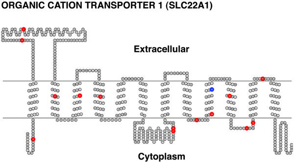
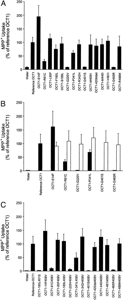
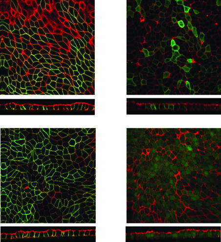
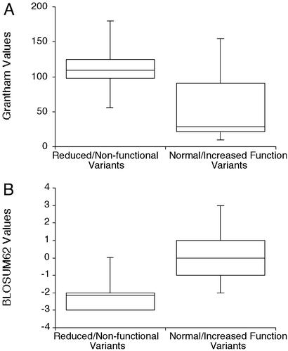
The organic cation transporter, OCT1, is a major hepatic transporter that mediates the uptake of many organic cations from the blood into the liver where the compounds may be metabolized or secreted into the bile. Because OCT1 interacts with a variety of structurally diverse organic cations, including clinically used drugs as well as toxic substances (e.g., N-methylpyridinium, MPP(+)), it is an important determinant of systemic exposure to many xenobiotics. To understand the genetic basis of extensive interindividual differences in xenobiotic disposition, we functionally characterized 15 protein-altering variants of the human liver organic cation transporter, OCT1, in Xenopus oocytes. All variants that reduced or eliminated function (OCT1-R61C, OCT1-P341L, OCT1-G220V, OCT1-G401S, and OCT1-G465R) altered evolutionarily conserved amino acid residues. In general, variants with decreased function had amino acid substitutions that resulted in more radical chemical changes (higher Grantham values) and were less evolutionarily favorable (lower blosum62 values) than variants that maintained function. A variant with increased function (OCT1-S14F) changed an amino acid residue such that the human protein matched the consensus of the OCT1 mammalian orthologs. Our results indicate that changes at evolutionarily conserved positions of OCT1 are strong predictors of decreased function and suggest that a combination of evolutionary conservation and chemical change might be a stronger predictor of function.


Figures

Figure 1
Figure 1
Secondary structure and alignment of OCT1 with coding region SNPs. The transmembrane topology diagram was rendered using the transmembrane protein display software TOPO [S. J. Johns (University of California, San Francisco) and R. C. Speth (Washington State University, Pullman), transmembrane protein display software available at the University of California, San Francisco Sequence Analysis Consulting Group web site, www.sacs.ucsf.edu/TOPO/topo.html]. Nonsynonymous amino acid changes are shown in red, and amino acid deletion is shown in blue.
Figure 2
Figure 2
Functional characterization of natural variants of OCT1. (A) Uptake of MPP+ in oocytes expressing the reference OCT1 and each of the 15 protein-altering OCT1 variants. (B) Recovery of function for six OCT1 variants with reduced or increased function. Dark-shaded columns represent uptake by variants; lighter-shaded columns represent uptake by variants changed to the OCT1 reference sequence at a single nucleotide position by site-directed mutagenesis. (C) Effect of double mutations on transporter function. X. laevis oocytes were injected with ≈50 ng of RNA. Uptake of MPP+ (0.9 μM unlabeled MPP+, 0.1 μM 3H-MPP+) was measured at room temperature after incubation for 1 h. Data are representative of experiments carried out with three different batches of oocytes. Each value represents mean ± SD from seven to nine oocytes.
Figure 3
Figure 3
Localization of GFP-tagged reference OCT1, OCT1-G465R, and OCT1-R488M in Madin–Darby canine kidney (MDCK) cells. MDCK cells were transfected with plasmids eGFP-OCT1, eGFP-OCT1-G465R, eGFP-OCT1-R488M, and eGFP, respectively. Stable clones were selected with G418. Cells were polarized by growth on filters for 7 days. The cells were fixed, permeabilized, stained for actin with Texas-red-conjugated X phalloidin, and visualized by confocal fluorescence microscopy. A horizontal section (Upper) and a vertical section with the apical membrane on top (Lower) are shown for each set of transfected cells. (Upper Left) eGFP-OCT1. (Upper Right) eGFP-OCT1-G465R. (Lower Left) eGFP-OCT1-R488M. (Lower Right) eGFP.
Figure 4
Figure 4
Grantham and BLOSUM62 values for OCT1 variants. (A) Grantham values for OCT1 variants with or without decreased function. Mean ± SE values for the variants with decreased function are 114 ± 20 versus 58.2 ± 19 for the variants with increased or normal function, respectively (P < 0.1). (B) BLOSUM62 values for OCT1 variants with or without decreased function. Mean ± SE values for the variants with decreased function are −2.2 ± 0.57 versus 0.0 ± 0.53 for the variants with increased or normal function, respectively (P < 0.02).

References

    1. Evans W E, Relling M V. Science. 1999;286:487–491. - PubMed
    1. Zhang L, Schaner M E, Giacomini K M. J Pharmacol Exp Ther. 1998;286:354–361. - PubMed
    1. Zhang L, Brett C M, Giacomini K M. Annu Rev Pharmacol Toxicol. 1998;38:431–460. - PubMed
    1. Zhang L, Dresser M J, Gray A T, Yost S C, Terashita S, Giacomini K M. Mol Pharmacol. 1997;51:913–921. - PubMed
    1. Gorboulev V, Ulzheimer J C, Akhoundova A, Ulzheimer-Teuber I, Karbach U, Quester S, Baumann C, Lang F, Busch A E, Koepsell H. DNA Cell Biol. 1997;16:871–881. - PubMed

Publication types

MeSH terms

Substances

LinkOut - more resources