. 2003 May;77(10):5547-56.
doi: 10.1128/jvi.77.10.5547-5556.2003.

Elimination of protease activity restores efficient virion production to a human immunodeficiency virus type 1 nucleocapsid deletion mutant

Affiliations

Elimination of protease activity restores efficient virion production to a human immunodeficiency virus type 1 nucleocapsid deletion mutant

David E Ott et al. J Virol. 2003 May.

Abstract

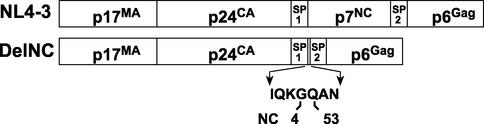
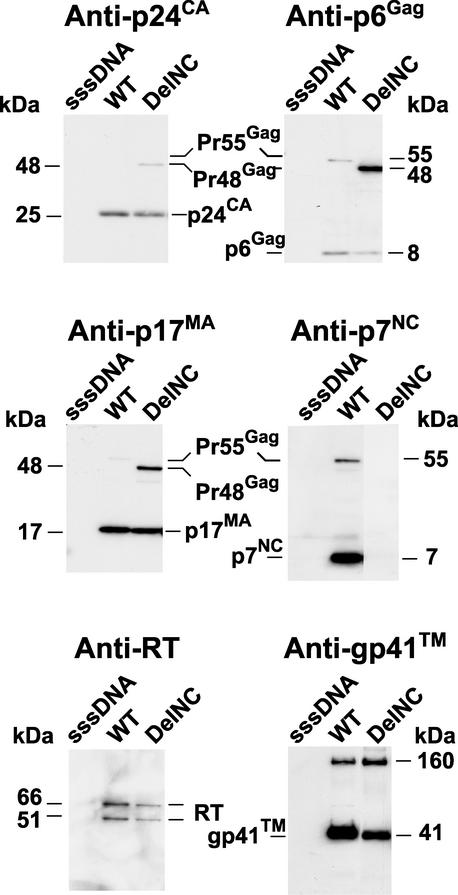
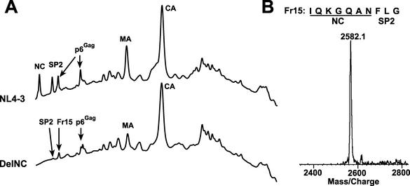
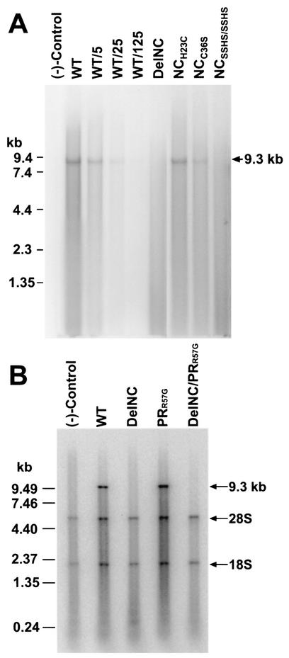
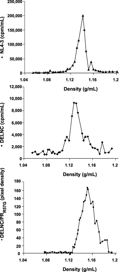
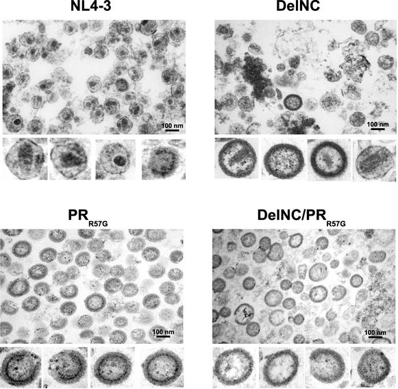
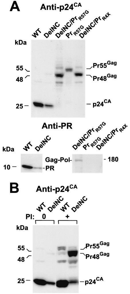
The nucleocapsid (NC) region of human immunodeficiency virus type 1 (HIV-1) Gag is required for specific genomic RNA packaging. To determine if NC is absolutely required for virion formation, we deleted all but seven amino acids from NC in a full-length NL4-3 proviral clone. This construct, DelNC, produced approximately four- to sixfold fewer virions than did the wild type, and these virions were noninfectious (less than 10(-6) relative to the wild type) and severely genomic RNA deficient. Immunoblot and high-pressure liquid chromatography analyses showed that all of the mature Gag proteins except NC were present in the mutant virion preparations, although there was a modest decrease in Gag processing. DelNC virions had lower densities and were more heterogeneous than wild-type particles, consistent with a defect in the interaction assembly or I domain. Electron microscopy showed that the DelNC virions displayed a variety of aberrant morphological forms. Inactivating the protease activity of DelNC by mutation or protease inhibitor treatment restored virion production to wild-type levels. DelNC-protease mutants formed immature-appearing particles that were as dense as wild-type virions without incorporating genomic RNA. Therefore, protease activity combined with the absence of NC causes the defect in DelNC virion production, suggesting that premature processing of Gag during assembly causes this effect. These results show that HIV-1 can form particles efficiently without NC.


Figures

FIG. 1.
FIG. 1.
DelNC mutant Gag. Diagrams of the Gag regions of the wild-type and DelNC constructs are presented. The NC sequences remaining after deletion of the majority of NC are displayed in single-amino-acid code just below the diagram, with the amino acid positions relative to NC at the fusion point indicated.
FIG. 2.
FIG. 2.
Immunoblots of DelNC virions. Immunoblots of virion preparations produced by transfection are presented. The antiserum or antibody used is indicated above the respective blots, and samples are identified above the respective lanes. Molecular masses, as calculated by relative mobility, and identities of bands are indicated at the margins of the blots. WT, wild type.
FIG. 3.
FIG. 3.
HPLC analysis of DelNC. (A) Chromatograms of equal percentages of NL4-3 and DelNC virus preparations are presented. Peaks containing Gag proteins are identified above the respective peaks. (B) Protein sequence analysis of fraction 15 is displayed at the top, with the DelNC amino acids underlined. MALDI mass spectrometry results for fraction 15 are presented at the bottom.
FIG. 4.
FIG. 4.
Analysis of particles for intact genomic RNA. (A) Northern blot analysis of virion RNA preparations. Samples contained 5.2 × 10⁶ cpm of RT activity from each mutant and wild-type (WT) virus preparation. The blot was probed with a 32P-labeled 8.1-kbp AvaI fragment from pNL4-3. In addition to the wild-type NL4-3 sample, 1/5, 1/25, and 1/125 dilutions of this sample were tested. Samples are identified above their respective lanes, with RNA markers indicated on the left and the size of the full-length genome indicated on the right. (B) Autoradiogram of metabolically 32P-labeled virions analyzed by RNA denaturing agarose gel electrophoresis. Virion preparations isolated from equal amounts of transfection culture supernatants were examined. Samplesare indicated at the top of the gel, RNA marker sizes are indicated on the left, and the positions of the 9.3-kb viral RNA and 28S and 18S ribosomal bands are indicated on the right. For both panels, “(−)-Control” denotes a virus preparation produced from transfected sssDNA.
FIG. 5.
FIG. 5.
Equilibrium density gradient centrifugation of viruses. Profiles of wild-type NL4-3, DelNC, and DelNC/PRR57G viruses subjected to centrifugation through 10 to 50% (wt/vol) sucrose gradients are presented. Amounts of virions detected as measured by RT activity ([3H]TMP incorporation) or by scanning densitometry (pixel gray-scale density) from a p6Gag immunoblot of selected fractions are reported on the y axis versus density of sucrose on the x axis. Virions analyzed are identified to the left of the respective graphs.
FIG. 6.
FIG. 6.
Electron micrographs of virions. Transmission electron micrographs of positively stained wild-type, DelNC, PRR57G, and DelNC/PRR57G virion preparations are presented at ×40,000 magnification. Enlarged images of representative virions present in this field and others are displayed underneath the field micrographs.
FIG. 7.
FIG. 7.
Immunoblots of protease-deficient virions. Immunoblots of virion preparations produced from transfection of various constructs (A) and those of virion preparations produced by transfection in the presence or absence of 10 nM saquinavir (B) are presented. The antiserum or antibody used is indicated above the respective blot, and the samples analyzed are identified above the respective lanes. Molecular masses, as calculated from relative mobility, and identities of bands are indicated at the margins of the blots. The addition of saquinavir in the production of virus samples is indicated as PI + above the appropriate lanes. WT, wild type.

References

    1. Accola, M. A., B. Strack, and H. G. Gottlinger. 2000. Efficient particle production by minimal Gag constructs which retain the carboxy-terminal domain of human immunodeficiency virus type 1 capsid-p2 and a late assembly domain. J. Virol. 74:5395-5402. - PMC - PubMed
    1. Adachi, A., S. Koenig, H. E. Gendelman, D. Daugherty, S. Gattoni-Celli, A. S. Fauci, and M. A. Martin. 1987. Productive, persistent infection of human colorectal cell lines with human immunodeficiency virus. J. Virol. 61:209-213. - PMC - PubMed
    1. Arrigo, S. J., and K. Huffman. 1995. Potent inhibition of human immunodeficiency virus type 1 (HIV-1) replication by inducible expression of HIV-1 PR multimers. J. Virol. 69:5988-5994. - PMC - PubMed
    1. Bennett, R. P., T. D. Nelle, and J. W. Wills. 1993. Functional chimeras of the Rous sarcoma virus and human immunodeficiency virus gag proteins. J. Virol. 67:6487-6498. - PMC - PubMed
    1. Berg, J. M., and Y. Shi. 1996. The galvanization of biology: a growing appreciation for the roles of zinc. Science 271:1081-1085. - PubMed

Publication types

MeSH terms

LinkOut - more resources