. 2003 May;77(10):5657-67.
doi: 10.1128/jvi.77.10.5657-5667.2003.

L-particle production during primary replication of pseudorabies virus in the nasal mucosa of swine

Affiliations

L-particle production during primary replication of pseudorabies virus in the nasal mucosa of swine

Nuria Alemañ et al. J Virol. 2003 May.

Abstract

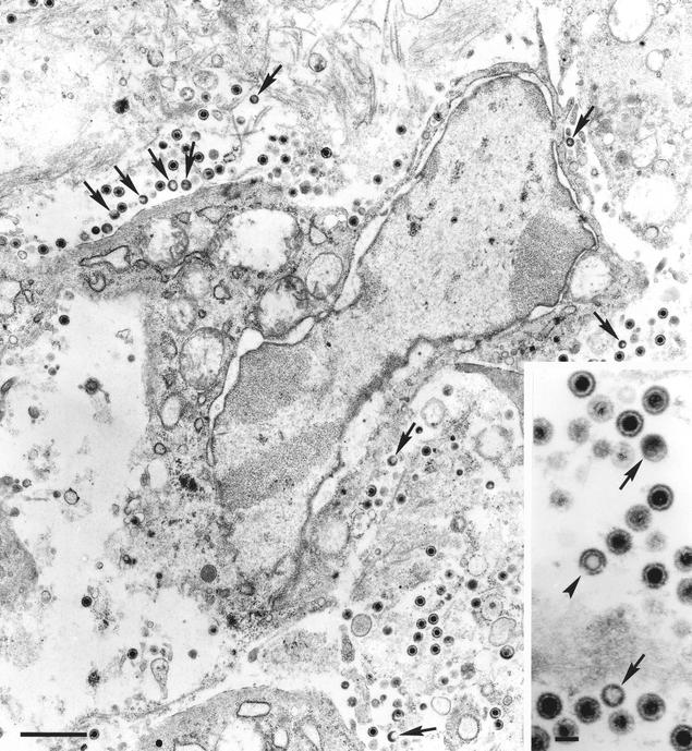
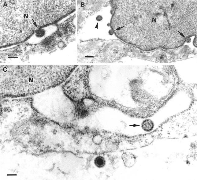
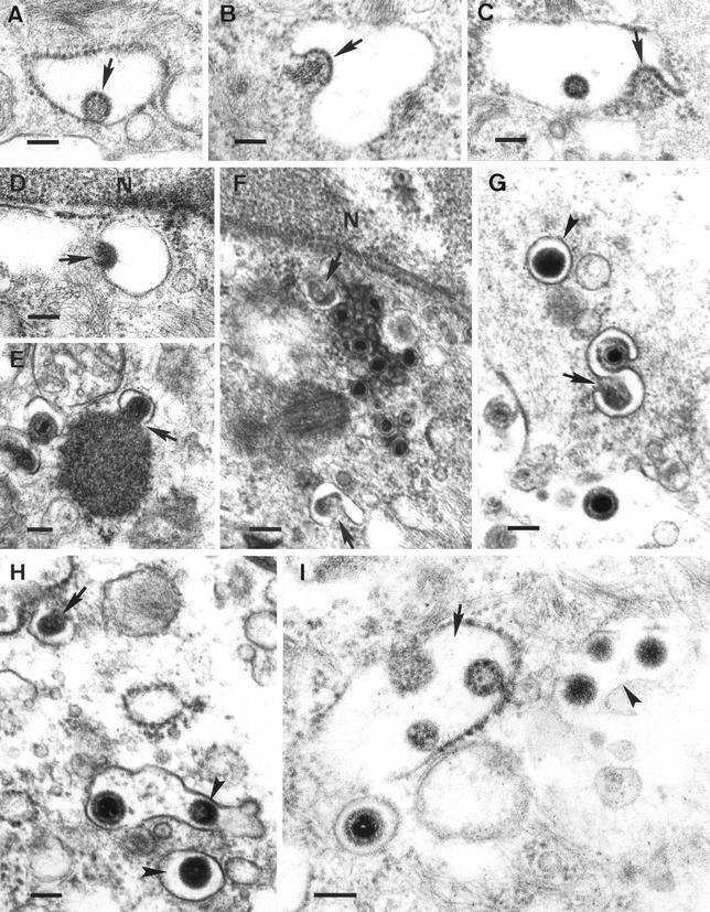
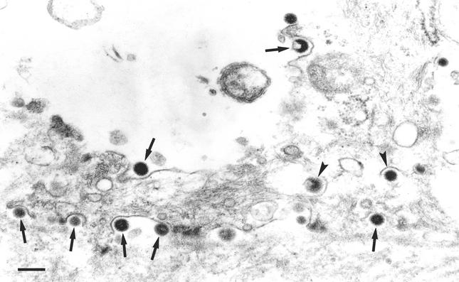
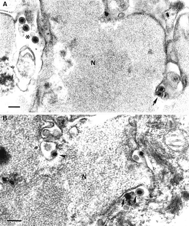
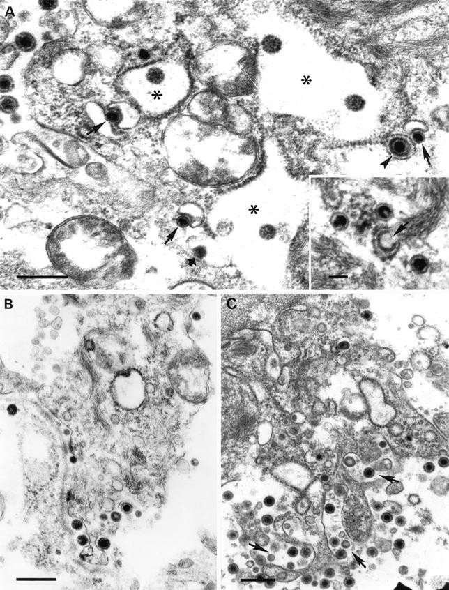
Different tissue culture cell lines infected with a number of alphaherpesviruses produce, in addition to virions, light particles (L particles). L particles are composed of the envelope and tegument components of the virion but totally lack the proteins of the capsid and the virus genome; therefore, they are noninfectious. In this electron microscopy report, we show that L particles are produced during primary replication of the alphaherpesvirus pseudorabies virus (PRV) in the nasal mucosa of experimentally infected swine, its natural host. Although PRV infected different types of cells of the respiratory and olfactory mucosae, PRV L particles were found to be produced exclusively by epithelial cells and fibroblasts. We observed that formation of noninfectious particles occurred by budding of condensed tegument at the inner nuclear membrane and at membranes of cytoplasmic vesicles, resulting in intracisternal and intravesicular L particles, respectively. Both forms of capsidless particles were clearly distinguishable by the presence of prominent surface projections on the envelope and the higher electron density of the tegument, morphological features which were only observed in intravesicular L particles. Moreover, intravesicular but not intracisternal L particles were found to be released by exocytosis and were also identified extracellularly. Comparative analysis between PRV virion and L-particle morphogenesis indicates that both types of virus particles share a common intracellular pathway of assembly and egress but that they show different production patterns during the replication cycle of PRV.


Figures

FIG. 1.
FIG. 1.
Low-magnification TEM micrograph of the lamina propria of the porcine nasal mucosa 72 h after intranasal inoculation of PRV strain E-974. Extracellular L particles (arrows) and virions can be observed in the connective matrix that surrounds the infected fibroblast. The inset shows two L particles (arrows), numerous virions, and an empty enveloped capsid (arrowhead). Bars, 1 μm and 150 nm (inset).
FIG. 2.
FIG. 2.
Formation of intracisternal L particles and passage into the RER. Budding of condensed tegument at the inner nuclear membrane (arrows in panels A and B) resulted in perinuclear L particles (arrowhead in B). An L particle (arrow) within a distended RER profile continuous with the nuclear envelope is shown in panel C, as is an extracellular virion. Bars, 150 nm in panels A and C and 250 nm in panel B. N, nucleus.
FIG. 3.
FIG. 3.
(A to C) Fusion between the envelope of intracisternal L particles (arrows) and the RER membrane. (D to H) Formation of intravesicular L particles. Budding of condensed tegument into cytoplasmic vesicles (arrows) resulted in L particles inside exocytic vesicles (arrowheads). Note the simultaneous budding of condensed tegument and a nucleocapsid into the same vesicle (G) as well as the presence of an exocytic vesicle containing both a virion and an L particle (H). (I) Intracisternal (arrow) and intravesicular (arrowhead) L particles. Note also the presence of an intravesicular virion. Bars, 150 nm. N, nucleus.
FIG. 4.
FIG. 4.
Exocytosis of L particles. The release of L particles (arrows) similar to those present within cytoplasmic vesicles (arrowheads) can be observed at the basolateral membrane of an epithelial cell. Bar, 250 nm.
FIG. 5.
FIG. 5.
Egress of nucleocapsids from the nucleus. (A) Budding of nucleocapsids at the inner nuclear membrane (arrow) resulted in primary enveloped nucleocapsids in the perinuclear cisterna (asterisk). (B) Direct fusion of the primary envelope with the outer nuclear membrane (arrows) resulted in release of naked nucleocapsids into the cytoplasm. Note also the primary envelopment of a nucleocapsid at the inner nuclear membrane (arrowhead). Bars, 250 nm. N, nucleus.
FIG. 6.
FIG. 6.
Secondary envelopment of nucleocapsids and exocytosis of virions. (A) Budding of intracytoplasmic nucleocapsids into cytoplasmic vesicles (arrows) resulted in mature virions inside exocytic vesicles (arrowhead). Note the presence of L particles within distended RER profiles (asterisks) and the formation of an intravesicular L particle (short arrow). The inset shows naked intracytoplasmic nucleocapsids adjacent to a RER profile marked with electron-dense granules (arrow) similar to those found after completion of the fusion event between the envelope of intracisternal L particles and the organelle membrane. (B and C) Simultaneous egress of virions and L particles at the basolateral membranes of epithelial cells. (C) Exocytosis of vesicles containing both virions and L particles (arrows). Bars, 500 nm and 100 nm (inset in panel A).

References

    1. Alconada, A., U. Bauer, B. Sodeik, and B. Hoflack. 1999. Intracellular traffic of herpes simplex virus glycoprotein gE: characterization of the sorting signals required for its trans-Golgi network localization. J. Virol. 73:377-387. - PMC - PubMed
    1. Alemañ, N., M. I. Quiroga, M. López-Peña, S. Vázquez, F. H. Guerrero, and J. M. Nieto. 2001. Induction and inhibition of apoptosis by pseudorabies virus in the trigeminal ganglion during acute infection of swine. J. Virol. 75:469-479. - PMC - PubMed
    1. Bates, P. A., and N. A. DeLuca. 1998. The polyserine tract of herpes simplex virus ICP4 is required for normal viral gene expression and growth in murine trigeminal ganglia. J. Virol. 72:7115-7124. - PMC - PubMed
    1. Browne, H., S. Bell, T. Minson, and D. W. Wilson. 1996. An endoplasmic reticulum-retained herpes simplex virus glycoprotein H is absent from secreted virions: evidence for reenvelopment during egress. J. Virol. 70:4311-4316. - PMC - PubMed
    1. Card, J. P., L. Rinaman, R. B. Lynn, B.-H. Lee, R. P. Meade, R. R. Miselis, and L. W. Enquist. 1993. Pseudorabies virus infection of the rat central nervous system: ultrastructural characterization of viral replication, transport, and pathogenesis. J. Neurosci. 13:2515-2539. - PMC - PubMed

Publication types

MeSH terms

Substances

LinkOut - more resources