. 2003 May;77(10):5829-36.
doi: 10.1128/jvi.77.10.5829-5836.2003.

Human immunodeficiency virus type 1 Env with an intersubunit disulfide bond engages coreceptors but requires bond reduction after engagement to induce fusion

Affiliations

Human immunodeficiency virus type 1 Env with an intersubunit disulfide bond engages coreceptors but requires bond reduction after engagement to induce fusion

L G Abrahamyan et al. J Virol. 2003 May.

Abstract

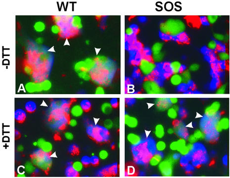
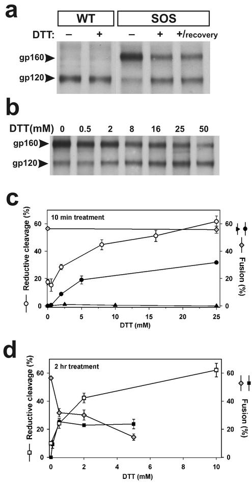
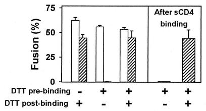
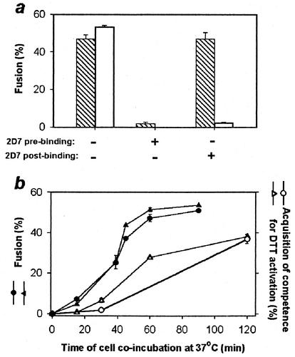
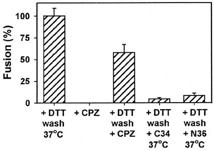
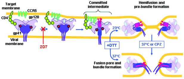
A mutant human immunodeficiency virus (HIV) envelope protein (Env) with an engineered disulfide bond between the gp120 and gp41 subunits (SOS-Env) was expressed on cell surfaces. With the disulfide bond intact, these cells did not fuse to target cells expressing CD4 and CCR5, but the fusion process did advance to an intermediate state: cleaving the disulfide bond with a reducing agent after but not before binding to target cells allowed fusion to occur. Through the use of an antibody directed against CCR5, it was found that at the intermediate stage, SOS-Env had associated with coreceptors. Reducing the disulfide bond after this intermediate had been reached resulted in hemifusion at low temperature and fusion at physiological temperature. The addition of C34 or N36, peptides that prevent six-helix bundle formation, at the hemifused state blocked the fusion that would have resulted after raising the temperature. Thus, Env has not yet folded into six-helix bundles after hemifusion has been achieved. Because SOS-Env binds CCR5, it is suggested that the conformational changes in wild-type Env that result from this binding cause disengagement of gp120 from gp41 in the region of the engineered bond. It is proposed that this disengagement is the event that directly frees gp41 to undergo the conformational changes that lead to fusion. The intermediate state achieved prior to reduction of the disulfide bond was stable. The capture of this configuration of Env could yield a suitable antigen for vaccine development, and it may also be a target for pharmacological intervention against HIV-1 entry.


Figures

FIG. 1.
FIG. 1.
Cell-cell fusion induced by WT-Env (left panels) and SOS-Env (right panels). Effector cells expressing WT-Env or SOS-Env were loaded with calcein (green), and target cells were loaded with CMAC (blue) and colabeled with the membrane dye DiI (red). Cell fusion was examined after coincubation for 2 h at 37°C in the absence (A and B) or in the presence (C and D) of 0.5 mM DTT. Fused cells are marked by arrowheads.
FIG. 2.
FIG. 2.
Reductive cleavage of SS-bond and fusion activity of SOS-Env. (a) Wild-type (lanes 1 and 2) and SOS (lanes 3 to 5), treated or not treated with 25 mM DTT (10 min at 37°C), were assayed either immediately after reduction (lanes 1 to 4) or after a 2-h incubation in phosphate-buffered saline (lane 5). (b) Dose dependence of the reductive cleavage of SS-bonds by DTT. (c) The extent of reductive cleavage of SOS as a function of DTT concentration determined from gels (as in b) as the ratio of total gp120 to the sum of gp120 and gp160 (open circles). In parallel experiments, the extent of fusion was quantified for SOS cells treated with DTT either before (solid triangles) or after (solid circles) incubation with target cells. The extent of fusion after cells expressing WT-Env were exposed to DTT for 10 min was the same as in the absence of the exposure (gray diamonds). (d) The extent of fusion between cells for WT-Env (gray diamonds) and SOS-Env (solid squares) as a function of DTT concentration continuously maintained for 2 h. The increase in the extent of SS-bond reduction with DTT concentration is shown (open squares).
FIG. 3.
FIG. 3.
SOS-Env is activated by DTT applied after but not before binding to target cells for 1 h at 37°C. The extent of fusion induced by SOS-Env (hatched bars) and WT-Env (open bars) is shown for different DTT treatment protocols. In all cases, 25 mM DTT was maintained for 10 min at 37°C. In separate experiments, SOS cells were incubated with 25 μg of sCD4 per ml before the SS-bond was reduced with DTT (last two bars).
FIG. 4.
FIG. 4.
DTT activates SOS-Env after SOS-Env engages coreceptor of the target cell. (a) Extent of fusion induced by SOS-Env (cross-hatched bars) and by WT-Env (open bars) after the inhibitory 2D7 antibody was added to SOS cells and target cells either pre- or postbinding. (b) Kinetics of wild-type Env-induced fusion (solid symbols) and formation of a DTT-activatable state as a function of time of coincubating SOS cells with target cells (open symbols). Cells expressing SOS-Env and WT-Env were either treated (open and solid circles, respectively) or not treated (open and solid triangles, respectively) with DTT prior to incubation with target cells.
FIG. 5.
FIG. 5.
Characterization of fusion intermediate arrested by SS-bonds. SOS and target cells arrested at the committed state were exposed to 0.5 mM CPZ either before (second bar) or after (third bar) DTT treatment. Alternatively, cells captured at the committed state and treated with DTT were allowed to bind the C34 (0.4 μM) or N36 (25 μM) peptide and then brought back to the optimal temperature (fourth and fifth bars, respectively).
FIG. 6.
FIG. 6.
Model for membrane fusion mediated by SOS-Env. The SS-bonds between gp120 (light purple) and gp41 (dark purple) of SOS-Env are shown by red starbursts. CD4 is green, and CCR5 is dark cyan. The fusion peptides (red arrows) insert into the target cell membrane at the committed state. The amino-terminal trimeric coiled coil of gp41 is shown as three abutted red cylinders, and the carboxy-terminal heptad repeat regions are shown by blue cylinders. The gp120 subunits, after disengagement from gp41, are shown as being paler than before disengagement. Reduction of the SS-bonds allows gp41 to fold into a prebundle conformation and induce hemifusion at a temperature that is not permissive for fusion or to fold into a six-helix bundle and induce fusion pore formation at the optimal temperature.

References

    1. Allan, J. S., J. E. Coligan, F. Barin, M. F. McLane, J. G. Sodroski, C. A. Rosen, W. A. Haseltine, T. H. Lee, and M. Essex. 1985. Major glycoprotein antigens that induce antibodies in AIDS patients are encoded by HTLV-III. Science 228:1091-1094. - PubMed
    1. Barnett, A. L., and J. M. Cunningham. 2001. Receptor binding transforms the surface subunit of the mammalian C-type retrovirus envelope protein from an inhibitor to an activator of fusion. J. Virol. 75:9096-9105. - PMC - PubMed
    1. Berger, E. A., P. M. Murphy, and J. M. Farber. 1999. Chemokine receptors as HIV-1 coreceptors: roles in viral entry, tropism, and disease. Annu. Rev. Immunol. 17:657-700. - PubMed
    1. Binley, J. M., R. W. Sanders, B. Clas, N. Schuelke, A. Master, Y. Guo, F. Kajumo, D. J. Anselma, P. J. Maddon, W. C. Olson, and J. P. Moore. 2000. A recombinant human immunodeficiency virus type 1 envelope glycoprotein complex stabilized by an intermolecular disulfide bond between the gp120 and gp41 subunits is an antigenic mimic of the trimeric virion-associated structure. J. Virol. 74:627-643. - PMC - PubMed
    1. Binley, J. M., C. S. Cayanan, C. Wiley, N. Schülke, W. C. Olson, and D. R. Burton. 2003. Redox-triggered infection by disulfide-shackled human immunodeficiency virus type 1 pseudovirions. J. Virol. 77:5678-5684. - PMC - PubMed

Publication types

MeSH terms