. 2003 May;77(10):5964-74.
doi: 10.1128/jvi.77.10.5964-5974.2003.

Differentiation of varicella-zoster virus ORF47 protein kinase and IE62 protein binding domains and their contributions to replication in human skin xenografts in the SCID-hu mouse

Affiliations

Differentiation of varicella-zoster virus ORF47 protein kinase and IE62 protein binding domains and their contributions to replication in human skin xenografts in the SCID-hu mouse

Jaya Besser et al. J Virol. 2003 May.

Abstract

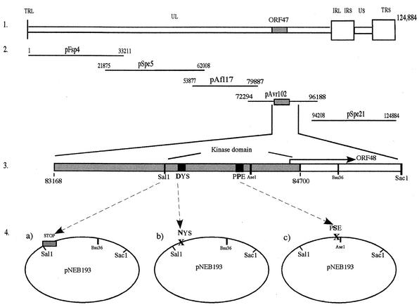
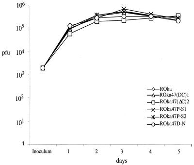
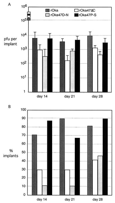
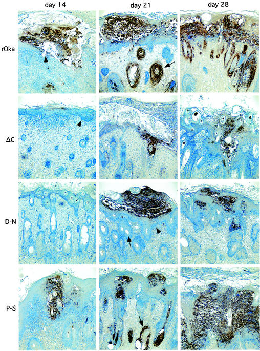
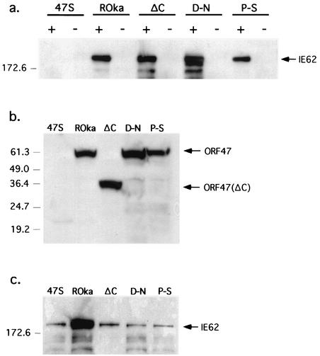
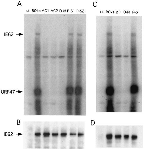
To investigate the role of the ORF47 protein kinase of varicella-zoster virus (VZV), we constructed VZV recombinants with targeted mutations in conserved motifs of ORF47 and a truncated ORF47 and characterized these mutants for replication, phosphorylation, and protein-protein interactions in vitro and for infectivity in human skin xenografts in the SCID-hu mouse model in vivo. Previous experiments showed that ROka47S, a null mutant that makes no ORF47 protein, did not replicate in skin in vivo (J. F. Moffat, L. Zerboni, M. H. Sommer, T. C. Heineman, J. I. Cohen, H. Kaneshima, and A. M. Arvin, Proc. Natl. Acad. Sci. USA 95:11969-11974, 1998). The construction of VZV recombinants with targeted ORF47 mutations made it possible to assess the effects on VZV infection of human skin xenografts of selectively abolishing ORF47 protein kinase activity. ORF47 mutations that resulted in a C-terminal truncation or disrupted the DYS kinase motif eliminated ORF47 kinase activity and were associated with extensive nuclear retention of ORF47 and IE62 proteins in vitro. Disrupting ORF47 kinase function also resulted in a marked decrease in VZV replication and cutaneous lesion formation in skin xenografts in vivo. However, infectivity in vivo was not blocked completely as long as the capacity of ORF47 protein to bind IE62 protein was preserved, a function that we identified and mapped to the N-terminal domain of ORF47 protein. These experiments indicate that ORF47 kinase activity is of critical importance for VZV infection and cell-cell spread in human skin in vivo but suggest that it is the formation of complexes between ORF47 and IE62 proteins, both VZV tegument components, that constitutes the essential contribution of ORF47 protein to VZV replication in vivo.


Figures

FIG. 1.
FIG. 1.
Schematic illustration of the construction of ORF47 mutants. (Panel 1) VZV genome, showing the localization of the ORF47 gene. (Panel 2) Five-cosmid system used to produce mutant viruses. ORF47 is located in pAvr102. (Panel 3) Schematic depiction of ORF47 indicating the coding sequence of the kinase domain in the 3′ half of the gene and the conserved kinase motifs that were targets for point mutations (shown in amino acid single-letter code). (Panel 4) A 2.5-kb SalI-SacI fragment subcloned into pNEB193 (via a 12-kb SalI-AscI fragment cloned into pNEB193; step not shown) containing the 3′ half of ORF47 and downstream sequences. This plasmid was used to produce the ORF47 mutations as follows: (a) insertion of STOP codons at the beginning of the kinase domain code, yielding the ORF47ΔC construct; (b) point mutation in the DYS motif, yielding the ORF47D-N construct; and (c) point mutation in the PPE motif, yielding the ORF47P-S construct. All mutants were constructed via PCR using the SalI restriction site as a sense and mutagenesis primer and a Bsu36I site 1,014 bp downstream of SalI as a location for the antisense primer (a and b) or an AseI site located 250 bp downstream of the SalI site and very close to the PPE motif as a location for the antisense and mutagenesis primer (c). All mutations were reinserted into pAvr102 via the SalI-AscI construct.
FIG. 2.
FIG. 2.
Replication of VZV in melanoma cells. Melanoma cells were inoculated on day 0 with 2 × 10³ PFU of rOka or rOka mutants. Two rOka47ΔC and rOka47P-S mutants were generated and tested independently. Aliquots were harvested daily for 5 days, and infectious foci were determined by titration on melanoma cell monolayers. Each time point represents the mean of results for at least three wells.
FIG. 3.
FIG. 3.
Replication of VZV ORF47 mutants in skin xenografts in SCID-hu mice. Skin tissue was infected with rOka, rOka47ΔC, rOka47D-N, and rOka47P-S grown in HEL cells. (A) Samples were harvested after 14, 21, and 28 days, and virus was titrated on melanoma monolayers. Each time point represents the mean number of plaques from two experiments, with four to six implants per experiment. Implants that did not contain infectious virus were excluded from the average. Inocula (shown as boxes on the y axis) are given as PFU/10 μl, which represents the amount used to infect each implant. (B) Percentages of implants that contained virus. The average of all implants in two experiments was computed. A total of 28 implants were infected with rOka, 31 implants were infected with rOka47ΔC, 32 implants were infected with rOka47D-N, and 30 implants were infected with rOka47P-S.
FIG.4.
FIG.4.
Immunohistologic staining of skin xenografts. Paraffin sections of xenografts collected at days 14, 21, and 28 were stained with human polyclonal VZV antiserum to visualize skin lesions (×10 magnification). Xenograft infected with rOka shows extended lesions, dissolution of the basement membrane layer (arrowhead), and infected hair follicles at days 21 (arrow) and 28. Xenografts infected with rOka47ΔC (ΔC) and rOka47D-N (D-N) show smaller lesions, the basement membrane remained intact (arrowheads, ΔC [day 14] and D-N [day 21]), and hair follicles were not infected (arrow, D-N). One single hair follicle shows infection with rOka47D-N at day 28. Xenografts infected with rOka47P-S (P-S) have larger lesions and show dissolution of the basement membrane. Multiple hair follicles were infected (arrow, P-S [day 21]).
FIG. 5.
FIG. 5.
Expression of CKII in cell cultures and SCID-hu skin xenografts. Lane 1, CKII α and β chain (Upstate Biotechnology, Inc.) (+); lane 2, melanoma cells (MeWo); lane 3, MRC-5 cells; lane 4, HEL cells; lane 5, skin xenograft cells. Lane 1 was used as positive control. All other lanes showed similar levels of CKII expression.
FIG. 6.
FIG. 6.
Immunoprecipitation of IE62 protein with ORF47 antiserum from melanoma cells infected with rOka or rOka ORF47 mutants. (a) Melanoma cells were infected with ROka47S (47S), rOka, rOka47ΔC (ΔC), rOka47D-N (D-N), or rOka47P-S (P-S). Infected cell lysates were incubated with ORF47 antiserum (+) or preimmune serum (−) and subjected to SDS-PAGE. A Western blot filter was probed with IE62 antiserum. A band slightly above the 172.6-kDa marker (which corresponds to the 175-kDa weight of IE62) was detected in lanes rOka, ΔC, D-N, and P-S (containing the ORF47 antiserum) but not in the lanes containing the ROka47S virus (kindly provided by J. Cohen, National Institutes of Health). (b and c) Western blot of total lysates. As a control, 20-μl portions of the infected cell lysate described above were directly subjected to SDS-PAGE. The filters were probed with ORF47 antiserum (b) or IE62 antiserum (c).
FIG. 7.
FIG. 7.
ORF47 protein kinase assay. (A) Melanoma cells were left uninfected (ui) or were infected with rOka, with both rOka47ΔC mutants (ΔC1 and ΔC2), with rOka47D-N (D-N), or with both rOka47P-S mutants (P-S1 and P-S2). A kinase assay was performed, lysates were subjected to SDS-PAGE, and proteins were transferred to membrane and autoradiographed. Phosphorylated bands of IE62 and ORF47 were determined by size. (B) After the radioactive signal decayed, the filter was probed with IE62 antiserum to show equal amounts of IE62 in all samples except uninfected cells. (C) rOka, rOka47ΔC, rOka47D-N, and rOka47P-S were cultured in melanoma cells after 28 days of growth in human skin xenografts in SCID-hu mice. Cell lysates were subjected to kinase assays as described above. (D) IE62 equal binding control.
FIG.8.
FIG.8.
Confocal analysis of melanoma cells infected with rOka and rOka ORF47 mutants for expression of ORF47 and IE62 proteins. Melanoma cells were infected with rOka, rOka47ΔC (ΔC), rOka47D-N (D-N), or rOka47P-S (P-S) for 30 h. ORF47 kinase was detected with a secondary fluorescein isothiocyanate-conjugated antibody (green), and IE62 was detected with a secondary Texas Red-conjugated antibody (red). Images of each row were merged. Colocalizations of ORF47 kinase and IE62 appear as yellow.

References

    1. Arvin, A. M. 2001. Varicella-zoster virus, p. 2731-2767. In B. N. Fields, D. M. Knipe, and P. M. Howley (ed.), Fields virology. Lippincott, Philadelphia, Pa.
    1. Bryant, D., and J. T. Parsons. 1983. Site-directed point mutation in the src gene of Rous sarcoma virus results in an inactive src gene product. J. Virol. 45:1211-1216. - PMC - PubMed
    1. Cohen, J. I., and S. E. Straus. 2001. Varicella-zoster virus and its replication, p. 2707-2730. In B. N. Fields, D. M. Knipe, and P. M. Howley (ed.), Fields virology. Lippincott, Philadelphia, Pa.
    1. Grose, C. 1981. Variation on a theme by Fenner: the pathogenesis of chickenpox. Pediatrics 68:735-737. - PubMed
    1. Hanks, S. K., and T. Hunter. 1995. Protein kinases 6. The eukaryotic protein kinase superfamily: kinase (catalytic) domain structure and classification. FASEB J. 9:576-596. - PubMed

Publication types

MeSH terms

Substances

LinkOut - more resources