. 2003 May 13;100(10):5986-90.
doi: 10.1073/pnas.1035999100. Epub 2003 Apr 29.

IL-17 production from activated T cells is required for the spontaneous development of destructive arthritis in mice deficient in IL-1 receptor antagonist

Affiliations

IL-17 production from activated T cells is required for the spontaneous development of destructive arthritis in mice deficient in IL-1 receptor antagonist

Susumu Nakae et al. Proc Natl Acad Sci U S A. .

Abstract

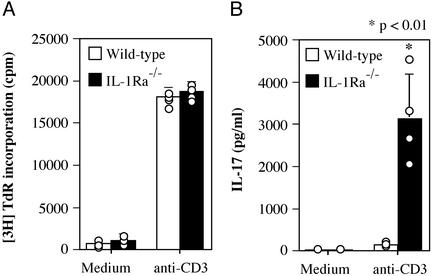
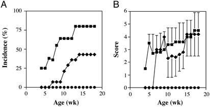
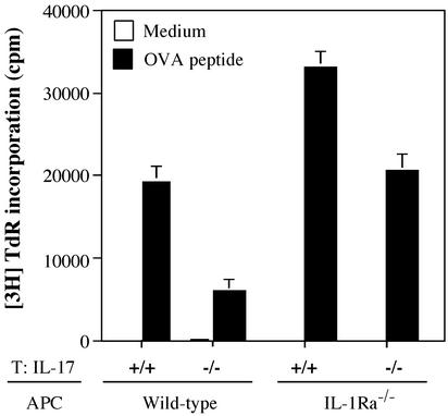
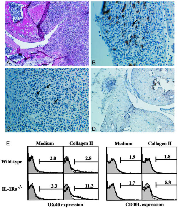
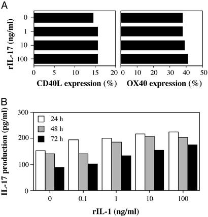
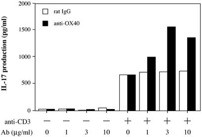
IL-17 is a T cell-derived, proinflammatory cytokine that is suspected to be involved in the development of various inflammatory diseases. Although there are elevated levels of IL-17 in synovial fluid of patients with rheumatoid arthritis, the pathogenic role of IL-17 in the development of rheumatoid arthritis remains to be elucidated. In this report, the effects of IL-17 deficiency were examined in IL-1 receptor antagonist-deficient (IL-1Ra(-/-)) mice that spontaneously develop an inflammatory and destructive arthritis due to unopposed excess IL-1 signaling. IL-17 expression is greatly enhanced in IL-1Ra(-/-) mice, suggesting that IL-17 activity is involved in the pathogenesis of arthritis in these mice. Indeed, the spontaneous development of arthritis did not occur in IL-1Ra(-/-) mice also deficient in IL-17. The proliferative response of ovalbumin-specific T cells from DO11.10 mice against ovalbumin cocultured with antigen-presenting cells from either IL-1Ra(-/-) mice or wild-type mice was reduced by IL-17 deficiency, indicating insufficient T cell activation. Cross-linking OX40, a cosignaling molecule on CD4(+) T cells that plays an important role in T cell antigen-presenting cell interaction, with anti-OX40 Ab accelerated the production of IL-17 induced by CD3 stimulation. Because OX40 is induced by IL-1 signaling, IL-17 induction is likely to be downstream of IL-1 through activation of OX40. These observations suggest that IL-17 plays a crucial role in T cell activation, downstream of IL-1, causing the development of autoimmune arthritis.


Figures

Figure 1
Figure 1
IL-17 production is markedly enhanced in T cells from IL-1Ra−/− mice. LN cells from wild-type and IL-1Ra−/− mice were cultured in the absence or presence of 1 μg/ml anti-CD3 mAb for 48 h. (A) Proliferative response was measured by [3H]thymidine incorporation. (B) IL-17 levels in the supernatants of A were determined by ELISA. Each circle represents an individual mouse, and an average and SD are shown. These results were reproducible in two independent experiments. The Student's t test was used for statistical evaluation of the results.
Figure 2
Figure 2
Development of arthritis in IL-1Ra−/− mice is completely suppressed by a deficiency of IL-17. (A) Incidence of arthritis that developed in IL-1Ra−/− mice on the BALB/c background. The incidences were compared among IL-17−/−, IL-17+/−, and IL-17+/+ mice. (B) Severity score of the mice. Average scores of the affected mice are shown and bars represent the SD. Squares, IL-17+/+ × IL-1Ra−/− (n = 10); diamonds, IL-17+/− × IL-1Ra−/− mice (n = 14); circles, IL-17−/− × IL-1Ra−/− mice (n = 10).
Figure 3
Figure 3
IL-17 is required for antigen-specific T cell priming. Proliferative responses of DO11.10 T cells against the ovalbumin 323–339 peptide in the presence of APCs from wild-type or IL-1Ra−/− mice were assessed by measuring the incorporation of [3H]thymidine after 3 days of culture. Effects of IL-17 deficiency were evaluated by using IL-17−/− DO11.10 T cells and IL-1Ra−/− APCs. Data shows an average ± SD of three wells. These results were reproducible in two independent experiments.
Figure 4
Figure 4
Enhanced OX40 and CD40L expression on T cells in IL-1Ra−/− mice. CD4+ and OX40+ cell infiltration was observed in synovial tissues of IL-1Ra−/− mice (AC) but not in wild-type mice (D). Both OX40 and CD40L expression on CD4+ T cells was enhanced in IL-1Ra−/− mice compared with wild-type mice after immunization with IIC (E). (A) Synovial tissues of IL-1Ra−/− mice; hematoxylin/eosin (H&E) staining (magnification, ×25). (B) Synovial tissues of IL-1Ra−/− mice stained with anti-CD4 mAb (magnification, ×100). Arrow shows positive cells. (C) Synovial tissues of IL-1Ra−/− mice stained with anti-OX40 mAb (magnification, ×100). Arrows show positive cells. (D) Synovial tissues of wild-type mice; H&E staining (magnification, ×25). (E) Seven days after immunization with IIC, LN cells were cultured in the absence or presence of 20 μg/ml IIC for 24 (CD40L expression) or 72 h (OX40 expression). Then, CD40L and OX40 expression in CD4+ population was analyzed by flow cytometry. Shaded area shows an isotype-matched control Ig staining.
Figure 5
Figure 5
IL-1 could not directly induce IL-17 in CD4+ T cells. (A) IL-17 could not induce CD40L and OX40 expression on CD4+ T cells. Splenic CD4+ T cells from wild-type mice were stimulated with plate-coated anti-mouse CD3 mAb (1 μg/ml) with or without rIL-17 for 24 (for CD40L expression) and 72 h (for OX40 expression). These results were reproducible in two independent experiments. (B) Splenic CD4+ T cells from wild-type mice were stimulated with or without rIL-1α together with plate-coated anti-mouse CD3 mAb for 24, 48, and 72 h. Then, IL-17 levels in culture supernatants were determined by ELISA. Data show a pooled supernatant from three wells. These results were reproducible in two independent experiments.
Figure 6
Figure 6
Cross-linking of OX40 on CD4+ T cells promotes IL-17 production. Cross-linking of OX40 could promote IL-17 production by CD4+ T cells. Splenic CD4+ T cells from wild-type mice were stimulated with plate-coated anti-mouse CD3 mAb (1 μg/ml) plus varying amounts of plate-coated anti-OX40 mAb or isotype control rat IgG for 96 h. Then, IL-17 levels in culture supernatants were determined by ELISA. Data show a pooled supernatant from three wells. These results were reproducible in three independent experiments.

References

    1. Yao Z, Painter S L, Fanslow W C, Ulrich D, Macduff B M, Spriggs M K, Armitage R J. J Immunol. 1995;155:5483–5486. - PubMed
    1. Fossiez F, Banchereau J, Murray R, Van Kooten C, Garrone P, Lebecque S. Int Rev Immunol. 1998;16:541–551. - PubMed
    1. Jovanovic D V, Di Battista J A, Martel-Pelletier J, Jolicoeur F C, He Y, Zhang M, Mineau F, Pelletier J P. J Immunol. 1998;160:3513–3521. - PubMed
    1. Shalom-Barak T, Quach J, Lotz M. J Biol Chem. 1998;273:27467–27473. - PubMed
    1. Kotake S, Udagawa N, Takahashi N, Matsuzaki K, Itoh K, Ishiyama S, Saito S, Inoue K, Kamatani N, Gillespie M T, et al. J Clin Invest. 1999;103:1345–1352. - PMC - PubMed

Publication types

MeSH terms