. 2003 May 13;100(10):6115-20.
doi: 10.1073/pnas.0937838100. Epub 2003 Apr 29.

Folding quality control in the export of proteins by the bacterial twin-arginine translocation pathway

Affiliations

Folding quality control in the export of proteins by the bacterial twin-arginine translocation pathway

Matthew P DeLisa et al. Proc Natl Acad Sci U S A. .

Abstract

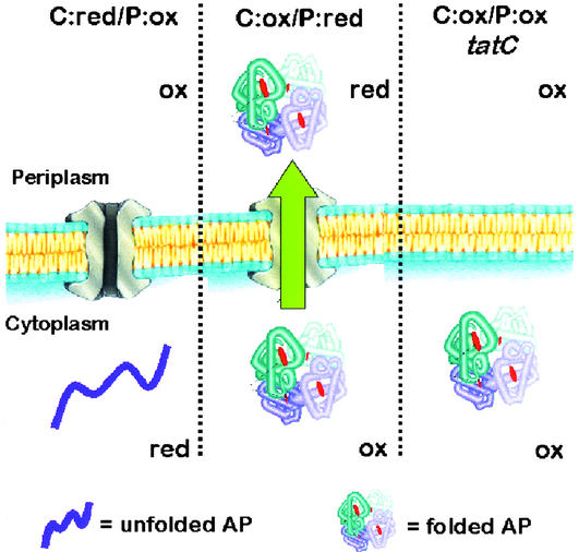
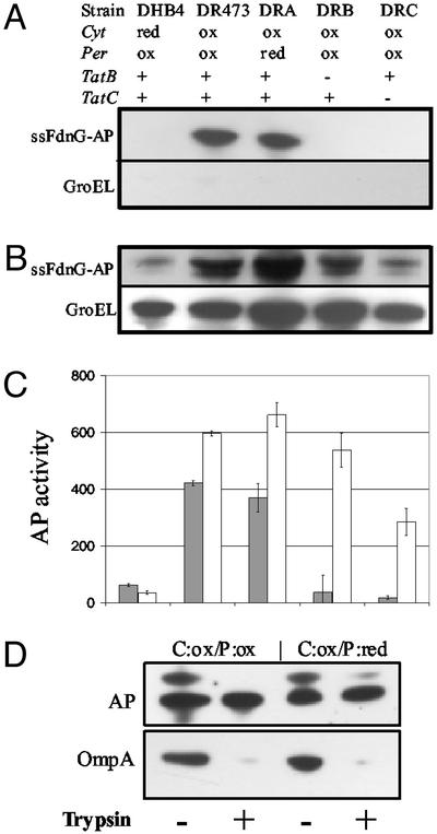
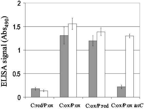
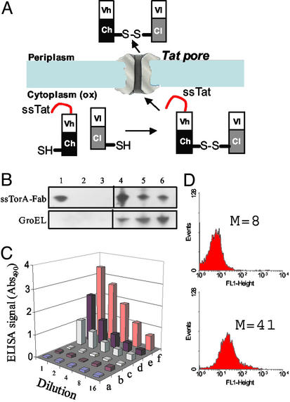
To examine the relationship between folding and export competence by the twin-arginine translocation (Tat) pathway we analyzed the subcellular localization of fusions between a set of eight putative Tat leader peptides and alkaline phosphatase in isogenic Escherichia coli strains that either allow or disfavor the formation of protein disulfide bonds in the cytoplasm. We show that export by the Tat translocator is observed only in strains that enable oxidative protein folding in the cytoplasm. Further, we show that other disulfide-containing proteins, namely single-chain Fv and heterodimeric F(AB) antibody fragments, are export-competent only in strains having an oxidizing cytoplasm. Functional, heterodimeric F(AB) protein was exported from the cytoplasm by means of a Tat leader peptide fused to the heavy chain alone, indicating that the formation of a disulfide-bonded dimer preceeds export. These results demonstrate that in vivo only proteins that have attained the native conformation are exported by the Tat translocator, indicating that a folding quality-control mechanism is intrinsic to the export process. The ability to export proteins with disulfide bonds and the folding proofing feature of the Tat pathway are of interest for biotechnology applications.


Figures

Figure 1
Figure 1
AP folding and export in different strain backgrounds. Ox and red refer to oxidizing and reducing redox potentials in the specified subcellular compartment. In C:ox (DR473) cells, AP is able to fold in the cytoplasm and thus can serve as a substrate for the Tat pathway. The localization of AP is not impaired in the C:ox/P:red strain DRA (DR473 dsbA). Deletion of tatC (or tatB) in the C:ox/P:ox strain blocks AP export.
Figure 2
Figure 2
Subcellular localization of ssFdnG-AP. Immunoblotting of periplasmic (A) and cytoplasmic (B) fractions from cells expressing ssFdnG-AP. Samples were normalized on the basis of the amount of total cell protein and resolved on SDS-12% polyacrylamide gels. GroEL was used as a fractionation marker by probing with anti-GroEL serum. (C) AP activity for the same periplasmic (filled bars) and cytoplasmic (open bars) fractions as in A and B. (D) Trypsin sensitivity analysis of periplasmic fractions collected from C:ox/P:ox and C:ox/P:red cells. Samples were separated on SDS-4–20% polyacrylamide gels and probed with anti-AP and anti-OmpA serum.
Figure 3
Figure 3
Tat export of a scFv. Detection of scFv by ELISA in the periplasmic and cytoplasmic fractions. Periplasmic (filled bars) and cytoplasmic (open bars) samples were serially diluted and started from the same amount of total protein. Data reported are from a 4-fold dilution and are the average of two independent experiments.
Figure 4
Figure 4
Tat export of an antidigoxin antibody fragment (FAB). (A) Tat export of FAB antibodies. (B) Western blotting of periplasmic (lanes 1–3) and cytoplasmic (lanes 4–6) fractions collected from cells coexpressing TorA-Fab fusion and ΔssDsbC. Strains used were (1, 4) C:ox/P:ox; (2, 5) C:ox/P:ox/tatC, and (3, 6) C:red/P:ox. All lanes were loaded with the same amount of total protein and anti-mouse IgG F(ab′)2 antibody was used to detect the FAB light chain. GroEL was used as a fractionation marker for the spheroplast fractions. (C) ELISA of periplasmic (a, c, and e) and cytoplasmic (b, d, and f) fractions collected from C:red/P:ox (a and b), C:ox/P:ox/tatC (c and d), and C:ox/P:ox (e and f) cells. (D) Flow cytometric analysis of C:red/P:ox cells (Upper) and C:ox/P:ox cells (Lower) cells expressing TorA-Fab and ΔssDsbC and labeled with FITC-digoxin.

References

    1. Pugsley A P. Microbiol Rev. 1993;57:50–108. - PMC - PubMed
    1. Stuart R A, Neupert W. Nature. 2000;406:575–577. - PubMed
    1. Settles A M, Yonetani A, Baron A, Bush D R, Cline K, Martienssen R. Science. 1997;278:1467–1470. - PubMed
    1. Weiner J H, Bilous P T, Shaw G M, Lubitz S P, Frost L, Thomas G H, Cole J A, Turner R J. Cell. 1998;93:93–101. - PubMed
    1. Cristobal S, de Gier J W, Nielsen H, von Heijne G. EMBO J. 1999;18:2982–2990. - PMC - PubMed

Publication types

Substances