. 2003 May 13;100(10):5795-800.
doi: 10.1073/pnas.0631579100. Epub 2003 Apr 29.

Resistance of cell membranes to different detergents

Affiliations

Resistance of cell membranes to different detergents

Sebastian Schuck et al. Proc Natl Acad Sci U S A. .

Abstract

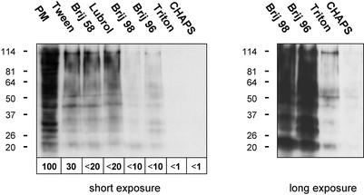
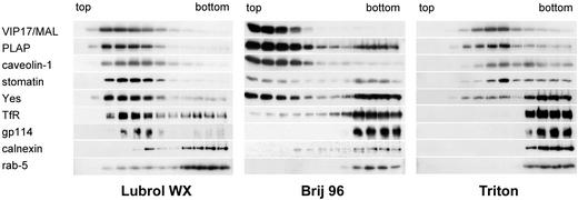
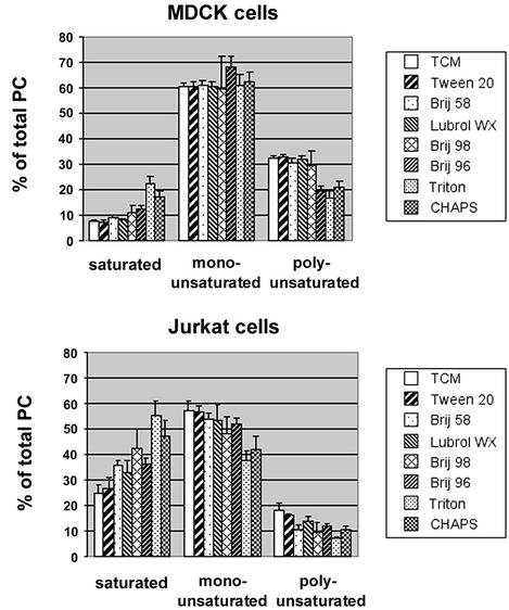
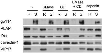
Partial resistance of cell membranes to solubilization with mild detergents and the analysis of isolated detergent-resistant membranes (DRMs) have been used operationally to define membrane domains. Given the multitude of detergents used for this purpose, we sought to investigate whether extraction with different detergents might reflect the same underlying principle of domain formation. We therefore compared the protein and lipid content of DRMs prepared with a variety of detergents from two cell lines. We found that the detergents differ considerably in their ability to selectively solubilize membrane proteins and to enrich sphingolipids and cholesterol over glycerophospholipids as well as saturated over unsaturated phosphatidylcholine. In addition, we observed cell type-dependent variations of the molecular characteristics of DRMs and the effectiveness of particular detergents. These results make it unlikely that different detergents reflect the same aspects of membrane organization and underscore both the structural complexity of cell membranes and the need for more sophisticated analytical tools to understand their architecture.


Figures

Figure 1
Figure 1
Protein content of plasma membrane DRMs from MDCK cells. Surface biotinylated MDCK cells were extracted with 1% Tween 20, 1% Brij 58, 0.5% Lubrol WX, 1% Brij 98, 0.5% Brij 96, and 1% Triton or 4% CHAPS. DRMs were prepared by flotation on sucrose step gradients. Equivalent aliquots of the starting material before detergent treatment, which contained the total plasma membrane protein (PM), and the various DRMs were analyzed by Western blotting using peroxidase-conjugated extravidin to reveal biotinylated proteins. Two exposures of the same membrane are shown. The amounts of biotinylated protein were quantified, setting PM to 100%.
Figure 2
Figure 2
DRM association of marker proteins from MDCK cells. MDCK-PLAP cell homogenate was extracted with 0.5% Lubrol WX, 0.5% Brij 96, or 1% Triton, and floated on linear sucrose gradients. The distribution of marker proteins was analyzed by immunoblotting. The light fractions from the top of the gradients are on the left, the heavy bottom fractions on the right.
Figure 3
Figure 3
Lipid contents of DRMs from MDCK and Jurkat cells. Lipids from TCMs and DRMs from 14C- or 32P-labeled MDCK and Jurkat cells were analyzed by TLC (Upper, 14C-labeled lipids; Lower, 32P-labeled lipids). GluC/GalC/LacC, glucosyl/galactosyl/lactosyl ceramide; SM, sphingomyelin; Gb5, pentosylglucoside; PE, phosphatidylethanolamine; PI, phosphatidylinositol; PS, phosphatidylserine. Asterisks denote lipids not identified by comigration with lipid standards.
Figure 4
Figure 4
Lipid contents of DRMs from MDCK and Jurkat cells. Lipids from the TLC plates shown in Fig. 3 were quantified and grouped into sphingolipids (GalC + GluC + LacC + SM + Gb5 for MDCK cells, GluC + SM for Jurkat cells, intensities taken from 14C-labeled lipids), cholesterol and glycerophospholipids (PE + PI + PS + PC, intensities taken from 32P-labeled lipids). Sphingolipid/glycerophospholipid and cholesterol/glycerophospholipid ratios relative to the ratios for TCMs are shown. GPL, glycerophospholipids.
Figure 5
Figure 5
Saturation profile of PCs in DRMs from MDCK and Jurkat cells. TCMs and DRMs were obtained as before, lipids were extracted, and PC was analyzed by mass spectrometry. Percent fractions of each PC species were determined and grouped into saturated (30:0, 31:0, 32:0, 34:0), monounsaturated (31:1, 32:1, 33:1, 34:1, 36:1), and polyunsaturated (32:2, 33:2, 34:2, 35:2, 36:3, 36:2, 38:3) PC species (in x:y, x indicates the total number of carbon atoms of the fatty acid side chains; y indicates the number of double bonds). Error bars represent standard deviations of three measurements of the same samples.
Figure 6
Figure 6
Manipulation of DRM association. MDCK cells were left untreated or treated with SMase, CD, saponin, or first SMase and then CD. After extraction with 1% Triton and flotation on Optiprep step gradients, two fractions containing the detergent-resistant (R, resistant) and detergent-soluble (S, soluble) material were collected and analyzed by immunoblotting.

References

    1. Schroeder R, London E, Brown D A. Proc Natl Acad Sci USA. 1994;91:12130–12134. - PMC - PubMed
    1. Ahmed S N, Brown D A, London E. Biochemistry. 1997;36:10944–10953. - PubMed
    1. Dietrich C, Bagatolli L A, Volovyk Z N, Thompson N L, Levi M, Jacobson K, Gratton E. Biophys J. 2001;80:1417–1428. - PMC - PubMed
    1. Ipsen J H, Karlstrom G, Mouritsen O G, Wennerstrom H, Zuckermann M J. Biochim Biophys Acta. 1987;905:162–172. - PubMed
    1. Brown D A, London E. J Membr Biol. 1998;164:103–114. - PubMed

Publication types