. 2003;13(3):399-412.
doi: 10.1002/hipo.10089.

Mossy fiber plasticity and enhanced hippocampal excitability, without hippocampal cell loss or altered neurogenesis, in an animal model of prolonged febrile seizures

Affiliations

Mossy fiber plasticity and enhanced hippocampal excitability, without hippocampal cell loss or altered neurogenesis, in an animal model of prolonged febrile seizures

Roland A Bender et al. Hippocampus. 2003.

Abstract

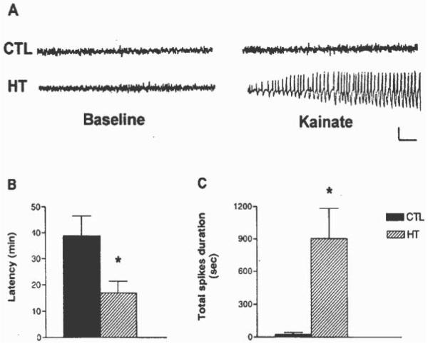
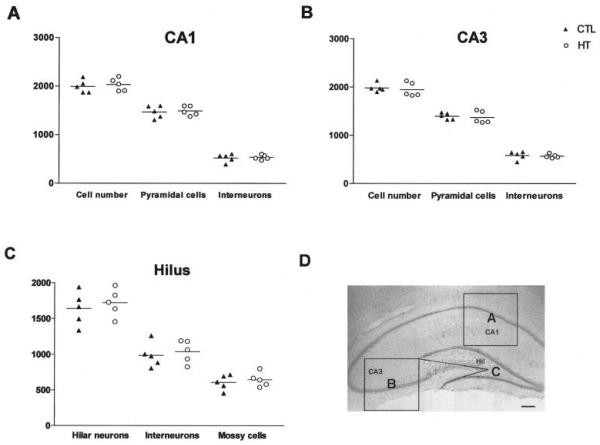
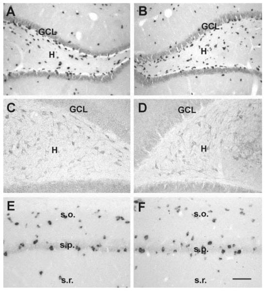
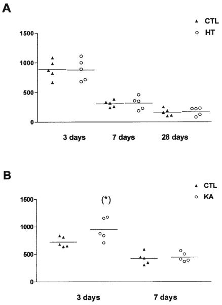
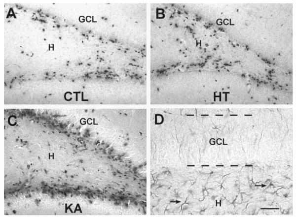
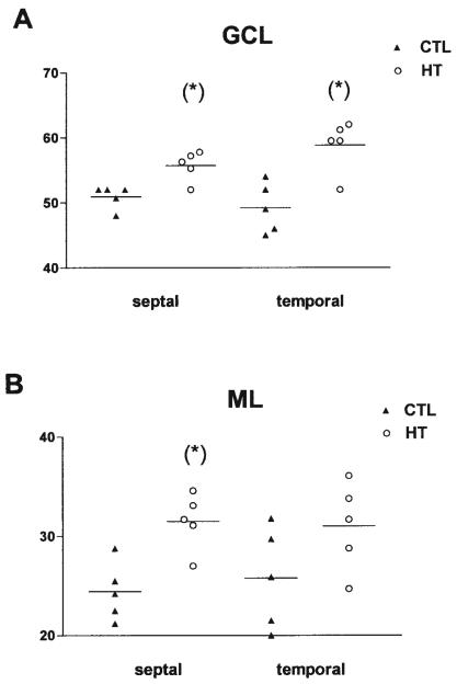
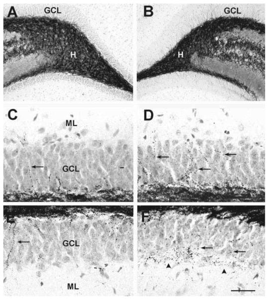
Seizures induced by fever (febrile seizures) are the most frequent seizures affecting infants and children; however, their impact on the developing hippocampal formation is not completely understood. Such understanding is highly important because of the potential relationship of prolonged febrile seizures to temporal lobe epilepsy. Using an immature rat model, we have previously demonstrated that prolonged experimental febrile seizures render the hippocampus hyperexcitable throughout life. Here we examined whether (1) neuronal loss, (2) altered neurogenesis, or (3) mossy fiber sprouting, all implicated in epileptogenesis in both animal models and humans, were involved in the generation of a pro-epileptic, hyperexcitable hippocampus by these seizures. The results demonstrated that prolonged experimental febrile seizures did not result in appreciable loss of any vulnerable hippocampal cell population, though causing strikingly enhanced sensitivity to hippocampal excitants later in life. In addition, experimental febrile seizures on postnatal day 10 did not enhance proliferation of granule cells, whereas seizures generated by kainic acid during the same developmental age increased neurogenesis in the immature hippocampus. However, prolonged febrile seizures resulted in long-term axonal reorganization in the immature hippocampal formation: Mossy fiber densities in granule cell- and molecular layers were significantly increased by 3 months (but not 10 days) after the seizures. Thus, the data indicate that prolonged febrile seizures influence connectivity of the immature hippocampus long-term, and this process requires neither significant neuronal loss nor altered neurogenesis. In addition, the temporal course of the augmented mossy fiber invasion of the granule cell and molecular layers suggests that it is a consequence, rather than the cause, of the hyperexcitable hippocampal network resulting from these seizures.


Figures

FIGURE 1
FIGURE 1
A: Hippocampal tracings (EEGs) obtained from adult rats before and after administration of a low-dose of kainic acid (KA). The normal tracings on the left are those from control (CTL) rats, as well as from those that had experienced experimental febrile seizures early in life (HT). The tracings on the right were obtained after KA administration. While hippocampal EEG activity remained normal in most controls (only two developed short seizures), high amplitude epileptiform spike-wave trains, and associated behavioral seizures, occurred in the HT rats. Calibration: vertical, 1 mV; horizontal, 1 s. B: Differential latency to the onset of KA-induced seizures in adult rats, depending on their previous seizure history: KA led to prolonged seizures in all HT rats (n = 9), whereas only 2 of 8 controls developed brief seizures. The latencies to the development of seizures were significantly shorter in HT group in comparison with latencies to the rare seizures of the control group. Values depict means ±SEM; *Significant difference: P < 0.05). C: Quantitative analysis of KA-induced total spikes duration (total duration of spike-and-wave discharges per hour recording) in rats that had sustained experimental febrile seizures early in life compared with littermate controls. After KA administration, hippocampi of HT animals exhibited spike-and-wave discharges of significantly longer total duration, compared with the CTL group (means ±SEM; *P < 0.05). Note: recording lasted for 180 min, or for 60 min after onset of status epilepticus.
FIGURE 2
FIGURE 2
Comparison of neuronal numbers in defined regions of the hippocampal formation of five rats with developmental febrile seizures (HT) and five age-matched controls (CTL) 3 months after the seizures. Data represent cell counts pooled from analyses of five sections (left and right hippocampus = 10 analyses per rat and subfield). A: CA1. B: CA3. C: Hilus. Note that in all analyzed subfields, neuronal numbers were not significantly different between the HT and control groups. In addition, the proportional relationships of these subpopulations were preserved: interneurons constituted ~26% of the neuronal population in CA1, ~29% in CA3 and ~60% in the hilus, mossy cells constituted ~37% of hilar neurons in both the HT and the control group. D: Demonstration of the areas analyzed: CA1 (frame A), CA3 (frame B), and hilus (frame C). Scale bar = 250 μm in D.
FIGURE 3
FIGURE 3
Neuronal populations which are typically vulnerable to seizure-induced excitotoxicity are not reduced in rats studied 3 months after developmental febrile seizures (B,D,F) when compared with age-matched controls (A,C,E). A,B: Hilar γ-aminobutyric acid (GABA)ergic interneurons visualized using in situ hybridization (ISH) for GAD67 mRNA. C,D: Hilar mossy cells, demonstrated using immunocytochemistry for AMPA-receptor subunits GluR2/3. E,F: CA1 interneurons, visualized using GAD67-ISH. GCL, granule cell layer; H, hilus; s.o., stratum oriens; s.p., stratum pyramidale; s.r., stratum radiatum. Scale bars = 80 μm in A,B; 50 μ min C–F.
FIGURE 4
FIGURE 4
Quantitative analysis of 5-bromo-2′-deoxyuridine (BrdU)-immunoreactive nuclei in the granule cell (GC) layer after (A) experimental febrile seizures (HT) lasting ~20 min, or (B) kainic acid (KA)-induced status epilepticus lasting ~120 min. A: Rats were injected with BrdU at 3, 7, or 28 days after the febrile seizures and perfused 48 h later. Data represent BrdU-positive nuclei counted in five sections (left and right hippocampus = 10 analyses per rat). The numbers of BrdU-labeled cells declined with age, due to the established developmental reduction in GC neurogenesis. However, no differences were found between HT and control animals at any age or time point. B: Rats were injected with BrdU at 3 or 7 days after KA-induced seizures and were perfused 48 h later. In contrast to the HT-induced seizures, KA-induced seizures significantly increased the number of BrdU-labeled cells at the 3-day time point (asterisk). The seizure-induced neurogenesis was transient and was no longer evident when rats were sacrificed 1 week after the seizures.
FIGURE 5
FIGURE 5
Photomicrographs of the dentate gyrus of immature rats subjected to experimental seizures at P10 (HT, B), kainic acid (KA)-induced prolonged seizures (KA, C) compared with controls (CTL, A). All animals were injected with 5-bromo-2′-deoxyuridine (BrdU) on P13. Sections were processed for BrdU immunocytochemistry following the experimental procedures described in Fig. 4. The numbers of immunoreactive nuclei did not differ between the HT and control groups (see also Fig. 4A). Increased neurogenesis was evident after KA seizures. The analysis included only BrdU-immunopositive nuclei within or subjacent to the granule cell layer (GCL). These were probably not astrocytes, because GFAP immunocytochemistry (D) demonstrated the presence of the astrocyte marker only well within the hilus. Scale bars = 50 μm in A–C; 25 μminD.
FIGURE 6
FIGURE 6
Quantitative analysis of mossy fiber innervation of the granule cell (GCL, A) and molecular (ML, B) layers of the dentate gyrus in septal and temporal hippocampus (n = 5, each group). The hippocampal formation of naive 3-month-old rats (CTL) was compared with those from animals sustaining experimental febrile seizures early in life (HT). Sections were processed for Timm's stain as described in the methods, and all analyses were carried out without knowledge of treatment group. Significantly increased numbers of mossy fibers traversed the GCL in the HT animals (asterisks, A). More fibers also penetrated the ML in the septal (but not temporal) hippocampus (B). The y-axis denotes the numbers of fibers per linear millimeter (mm) of the suprapyramidal blade of the GCL.
FIGURE 7
FIGURE 7
Photomicrographs of the dentate gyrus in Timm-stained sections from adult rats subjected to experimental febrile seizures 3 months earlier (B,D,F), or from controls (A,C,E). Top: Low-magnification views, to demonstrate similar technical processing of samples. Middle: Suprapyramidal blade of the granule cell layer (GCL) from controls, C), or from experimental animals (D). Bottom: analogous set of photographs from the infrapyramidal blade (E,F). The increased numbers of Timm-stained chains of boutons (arrows) are clearly visible in the sections from the experimental group. In addition to increased fiber number, an almost continuous band of Timm-positive boutons (arrowheads) in the GCL- molecular layer (ML) boundary can be seen in experimental (F), but not in control rats (E). H, hilus. Scale bars = 150 μm in A,B; 25 μm in C–F.

Similar articles

Cited by

References

    1. Adams B, Lee M, Fahnestock M, Racine RJ. Long-term potentiation trains induce mossy fiber sprouting. Brain Res. 1997;775:193–197. - PubMed
    1. Altman J, Bayer SA. Migration and distribution of two populations of hippocampal granule cell precursors during the perinatal and postnatal periods. J Comp Neurol. 1990;301:365–381. - PubMed
    1. André V, Marescaux C, Nehlig A, Fritschy JM. Alterations of hippocampal GABAergic system contribute to development of spontaneous recurrent seizures in the lithium-pilocarpine model of temporal lobe epilepsy. Hippocampus. 2001;11:452–468. - PubMed
    1. Babb TL, Kupfer WR, Pretorius JK, Crandall PH, Levesque MF. Synaptic reorganization by mossy fibers in human epileptic fascia dentata. Neuroscience. 1991;42:351–363. - PubMed
    1. Baram TZ, Ribak CE. Peptide-induced infant status epilepticus causes neuronal death and synaptic reorganization. NeuroReport. 1995;6:277–280. - PMC - PubMed

Publication types

MeSH terms