

CD44 in rheumatoid arthritis
- PMID: 12723975
- PMCID: PMC165042
- DOI: 10.1186/ar746
CD44 in rheumatoid arthritis
Abstract
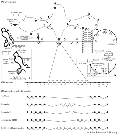
CD44 is a multistructural cell-surface glycoprotein that can theoretically generate close to 800 isoforms by differential alternative splicing. At present, several dozen isoforms are known. The polymorphic nature of CD44 might explain its multifunctionality and its ability to interact with many cell-surface and extracellular ligands, the principal one being hyaluronic acid (HA). Of the many CD44 functions, our review focuses on its involvement in cell-cell and cell-matrix interactions, as well as on its implication in the support of cell migration and the presentation of growth factors to their cognate receptors. Cells involved in pathological activities such as cancer cells and destructive inflammatory cells, and also normal cells engaged in physiological functions, use cell-surface CD44 for their localization and expansion at extravascular sites. This article reviews the evidence that the joint synovium of patients with rheumatoid arthritis (RA) contains considerable amounts of various CD44 isoforms as well as the HA ligand. The review also shows that anti-CD44 monoclonal antibody (mAb) directed against constant epitopes, shared by all CD44 isoforms, can markedly reduce the inflammatory activity of arthritis induced by collagen or proteoglycans in mice. Anti-CD44 mAb also interferes with the migration of RA synovial-like fibroblasts in vitro and is able to disturb the destructive interaction between RA synovial-like fibroblasts and the cartilaginous matrix. However, the transition from the experimental model to the patient's bedside is dependent on the ability to target the CD44 of cells engaged in RA pathology, while skipping the CD44 of normal cells.
Figures
References
-
- Lesley J, Hyman R, Kincade PW. CD44 and its interaction with extracellular matrix. Adv Immunol. 1993;54:271–335. - PubMed
-
- Naor D, Vogt Sionov R, Ish-Shalom D. CD44: structure, function and association with malignant process. Adv Cancer Res. 1997;71:241–319. - PubMed
-
- Lesley J, Hyman R. CD44 structure and function. Frontiers Biosci. 1998;3:d616–d630. - PubMed
-
- Naor D, Nedvetzki S, Golan I, Melnik L, Faitelson Y. CD44 in cancer. Crit Rev Clin Lab Sci. 2002;39:527–579. - PubMed
Publication types
MeSH terms
Substances
LinkOut - more resources
Full Text Sources
Other Literature Sources
Medical
Miscellaneous
