. 2003 May 1;548(Pt 3):823-36.
doi: 10.1113/jphysiol.2002.036772.

Receptor-coupled, DAG-gated Ca2+-permeable cationic channels in LNCaP human prostate cancer epithelial cells

Affiliations

Receptor-coupled, DAG-gated Ca2+-permeable cationic channels in LNCaP human prostate cancer epithelial cells

V Sydorenko et al. J Physiol. .

Abstract

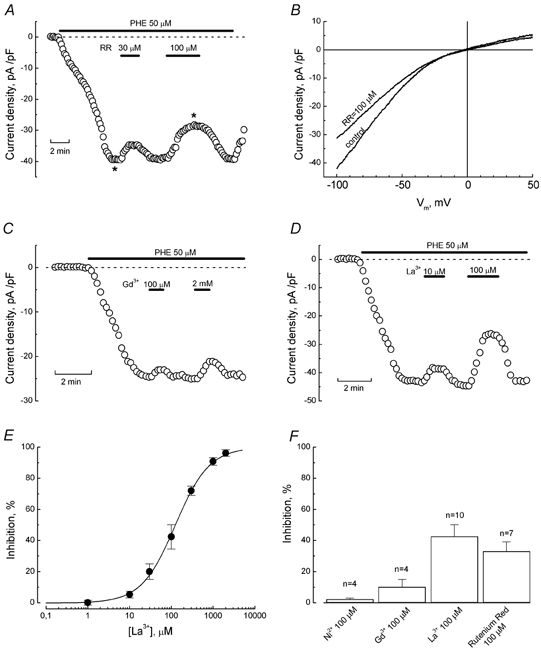
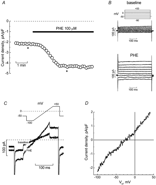
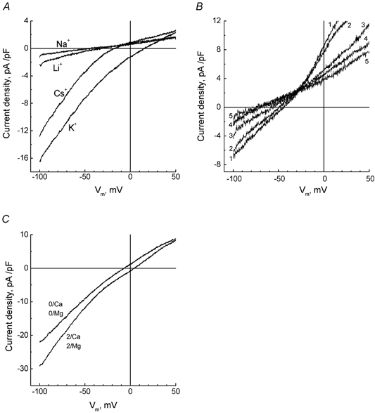
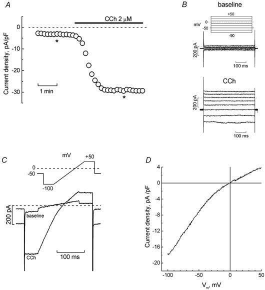
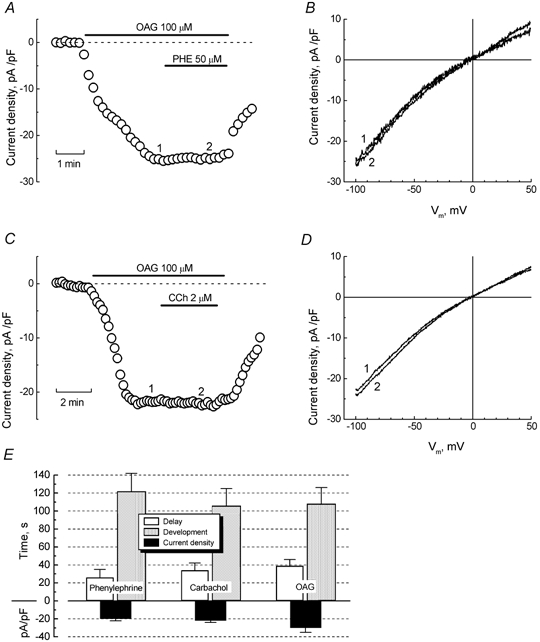
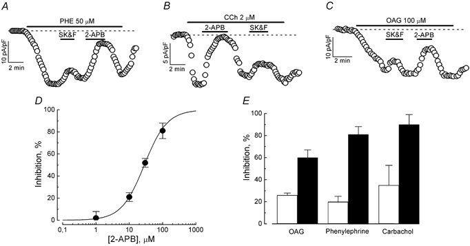
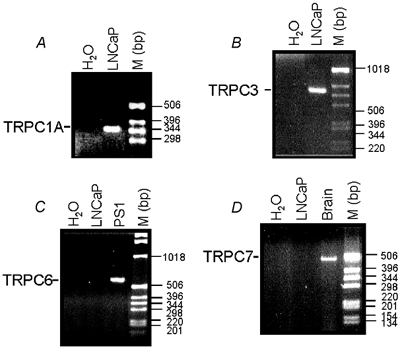
Although the prostate gland is a rich source of alpha1-adreno- (alpha1-AR) and m1-cholino receptors (m1-AChR), the membrane processes associated with their activation in glandular epithelial cells is poorly understood. We used the whole-cell patch-clamp technique to show that the agonists of the respective receptors, phenylephrine (PHE) and carbachol (CCh), activate cationic membrane currents in lymph node carcinoma of the prostate (LNCaP) human prostate cancer epithelial cells, which are not dependent on the filling status of intracellular IP3-sensitive Ca2+ stores, but directly gated by diacylglycerol (DAG), as evidenced by the ability of its membrane permeable analogue, OAG, to mimic the effects of the agonists. The underlying cationic channels are characterized by the weak field-strength Eisenman IV permeability sequence for monovalent cations (PK(25) > PCs(4.6) > PLi(1.4) > PNa(1.0)), and the following permeability sequence for divalent cations: PCa(1.0) > PMg(0.74) > PBa(0.6) > PSr(0.36) > PMn(0.3). They are 4.3 times more permeable to Ca2+ than Na+ and more sensitive to the inhibitor 2-APB than SK&F 96365. RT-PCR analysis shows that DAG-gated members of the transient receptor potential (TRP) channel family, including TRPC1 and TRPC3, are present in LNCaP cells. We conclude that, in prostate cancer epithelial cells, alpha1-ARs and m1-AChRs are functionally coupled to Ca2+-permeable DAG-gated cationic channels, for which TRPC1 and TRPC3 are the most likely candidates.


Figures

Figure 6
Figure 6. Sensitivity of PHE-evoked current in LNCaP cells to ruthenium red and polyvalent cations
A, time course of PHE-evoked inward current (at −100 mV) in a representative cell and its response to 30 μm and 100 μm ruthenium red (RR); interventions are marked with horizontal bars. B, ramp-derived I-V relationships of control PHE-induced current and PHE-induced current in the presence of 100 μm RR, acquired at times marked by asterisks on the time course in panel A. C and D, inhibition of PHE-induced current in representative cells by Gd3+ (100 μm and 2 mm, C) and La3+ (10 μm and 100 μm, D); interventions are marked with horizontal bars. E, dose-response relationship for the inhibitory action of La3+ on PHE-evoked current; data points - mean ±s.e.m., n = 4–10, smooth curve fit of the data points by Langmuir's isotherm; IC50 value is 124 μm. F, comparison of the inhibitory action (mean ±s.e.m.) of Ni2+ (100 μm), Gd3+ (100 μm), La3+ (100 μm), and ruthenium red (100 μm) on phenylephrine-evoked current.
Figure 1
Figure 1. Simulation of α 1-adrenoreceptors induces cationic current in LNCaP cells
A, time course of the development of inward current (measured at −100 mV) in a representative LNCaP cell bathed in normal extracellular solution, in response to phenylephrine (PHE, 100 μm, marked by horizontal bar). B, original baseline (top) and PHE-induced (bottom) currents recorded at the times marked by asterisks on the time course of panel A; pulse protocol is depicted above the baseline currents. C, superimposed baseline and PHE-induced currents from the same cell in response to the pulse containing a linear voltage-ramp portion (shown above the records). D, I-V relationship of PHE-induced current derived from ramps in panel C.
Figure 2
Figure 2. Selective properties of α1-AR-coupled membrane channels in LNCaP cells
A, ramp-derived I-V relationships of PHE-evoked current in a representative cell sequentially exposed to divalent cation-free extracellular solution containing 135 mm Na+, Cs+, Li+ or K+. B, same as in A, but for another cell sequentially exposed to NMDG-based extracellular solution containing 10 mm Ca2+ (1), Mg2+ (2), Ba2+ (3), Sr2+ (4) or Mn2+ (5). C, I-V relationships of PHE-evoked current in representative cell in standard Cs+-based extracellular solution (2/Ca, 2/Mg) and following omission of Ca2+ and Mg2+ (0/Ca, 0/Mg).
Figure 3
Figure 3. Muscarinic cholinoreceptor agonist, carbachol, induces cationic current in LNCaP cells similarly to PHE
A, development time course of inward current (measured at −100 mV) in the representative LNCaP cell in Cs+-based extracellular solution in response to carbachol (CCh, 2 μm, marked by horizontal bar). B, original baseline (top) and CCh-induced (bottom) currents recorded at the times marked by asterisks on the time course in panel A; pulse protocol is depicted above the baseline currents. C, superimposed baseline and CCh-induced currents from the same cell in response to the pulse containing a linear voltage-ramp portion (shown above the records). D, I-V relationship of CCh-induced current derived from ramps in panel C.
Figure 4
Figure 4. Stimulation of muscarinic and α1-adrenoreceptors converge on diacylglycerol-gated cationic channels
A, development time course of inward current (measured at −100 mV) in the representative LNCaP cell in Cs+-based extracellular solution in response to the membrane-permeable DAG analogue, 1-oleoyl-2-acetyl-sn-glycerol (OAG, 100 μm) alone, and following co-application with PHE (50 μm); interventions marked with horizontal bars. B, ramp-derived I-V relationships of OAG (1) and OAG + PHE (2) currents acquired at the respectively numbered times on the time course in panel A, showing virtually no difference in amplitude or reversal potential. C and D, same as in A and B, respectively, but with the interaction of OAG and CCh in another representative LNCaP cell. E, quantification of the delay, development periods, and maximal densities for PHE-, CCh- and OAG-induced currents (mean ±s.e.m., n = 10–15).
Figure 5
Figure 5. 2-APB and SK&F 96365 sensitivity of PHE-, CCh-, and OAG-evoked currents in LNCaP cells
A-C, time courses of PHE-, CCh-, and OAG-evoked inward currents (at −100 mV) in representative cells and responses to the application of 100 μm 2-APB and 25 μm SK&F 96365; interventions marked with horizontal bars. D, dose-response relationship for the inhibitory action of 2-APB on PHE-evoked current; data points - mean ±s.e.m., n = 4–7, smooth curve fit of the data points by Langmuir's isotherm; IC50 value is 28.2 μm. E, comparison of the inhibitory action of 100 μm 2-APB (▪) and 25 μm SK&F 96365 (□) on phenylephrine-, carbachol- and OAG-evoked currents (mean ±s.e.m., n = 4–7).
Figure 7
Figure 7. Expression of DAG-gated TRP members in LNCaP cells
RT-PCR analysis of the expression of human TRPC1A (A), TRPC3 (B), TRPC6 (C), and TRPC7 (D) transcripts in LNCaP cells. The expression products were obtained using the primers described in the Methods. Smooth muscle PS1 cells and brain tissue were used as positive controls for the detection of TRPC6 and TRPC7, respectively. M: DNA ladder.

References

    1. Berridge MJ. Calcium signalling and cell proliferation. Bioessays. 1995;17:491–500. - PubMed
    1. Berridge MJ, Bootman MD, Lipp P. Calcium - a life and death signal. Nature. 1998;935:645–648. - PubMed
    1. Brough GH, Wu S, Cioffi D, Moore TM, Li M, Dean N, Stevens T. Contribution of endogenously expressed Trp1 to a Ca2+-selective, store-operated Ca2+ entry pathway. FASEB J. 2001;15:1727–1738. - PubMed
    1. Braun FJ, Broad LM, Armstrong DL, Putney JW., Jr Stable activation of single Ca2+ release-activated Ca2+ channels in divalent cation-free solutions. J Biol Chem. 2001;276:1063–1070. - PubMed
    1. Brauner-Osborne H, Brann MR. Pharmacology of muscarinic acetylcholine receptor subtypes (m1-m5): high throughput assays in mammalian cells. Eur J Pharmacol. 1996;295:93–102. - PubMed

Publication types

MeSH terms

LinkOut - more resources