. 2003 May;23(10):3575-82.
doi: 10.1128/MCB.23.10.3575-3582.2003.

Mice lacking the giant protocadherin mFAT1 exhibit renal slit junction abnormalities and a partially penetrant cyclopia and anophthalmia phenotype

Affiliations

Mice lacking the giant protocadherin mFAT1 exhibit renal slit junction abnormalities and a partially penetrant cyclopia and anophthalmia phenotype

Lorenza Ciani et al. Mol Cell Biol. 2003 May.

Abstract

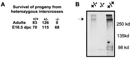
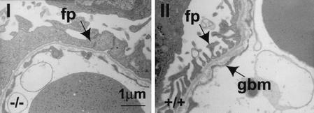
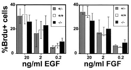
While roles in adhesion and morphogenesis have been documented for classical cadherins, the nonclassical cadherins are much less well understood. Here we have examined the functions of the giant protocadherin FAT by generating a transgenic mouse lacking mFAT1. These mice exhibit perinatal lethality, most probably caused by loss of the renal glomerular slit junctions and fusion of glomerular epithelial cell processes (podocytes). In addition, some mFAT1(-/-) mice show defects in forebrain development (holoprosencephaly) and failure of eye development (anophthalmia). In contrast to Drosophila, where FAT acts as a tumor suppressor gene, we found no evidence for abnormalities of proliferation in two tissues (skin and central nervous system [CNS]) containing stem and precursor cell populations and in which FAT is expressed strongly. Our results confirm a necessary role for FAT1 in the modified adhesion junctions of the renal glomerular epithelial cell and reveal hitherto unsuspected roles for FAT1 in CNS development.


Figures

FIG. 1.
FIG. 1.
(A) Schematic representation of the protocol used to mutate the mFAT1 locus, showing the wild-type (WT) locus (top), mFAT1 targeting vector (middle), and expected mutant allele after homologous recombination (bottom). A lacZ/Neo cassette was inserted in frame with the first ATG of the mFAT1 gene as described in Materials and Methods. The positive ES clones undergoing correct homologous recombination were identified by Southern blot analysis using a 3′ external probe and an internal probe on the lacZ gene. (B) Southern blot analysis (using the 3′ flanking probe) of EcoRI-digested tail genomic DNA from progeny of mFAT1 heterozygous intercrosses, showing the three possible genotypes obtained.
FIG. 2.
FIG. 2.
(A) The lacZ reporter gene was used to analyze the pattern of expression of the FAT1 gene in the embryo. Micrographs I to V show X-Gal staining in an intact (I) or sectioned (II to V) E12.5 embryo. The staining is strong in the forebrain (tel in panel I), mainly in the neocortex around the telencephalic vesicles (top part of panel II; note the lower level of staining in the ganglionic eminences in the lower part of the section), in the limb buds (panel I), in the developing eye (panel III), and in Rathke's pouch (panel IV). Panel V shows lacZ expression in the spinal cord of an E12.5 embryo. Note the staining in the region of the floor plate. e, eye; fp, floor plate; inf, infundibulum; lb, limb bud; ncx, neocortex; nl, neural layer; nc, notochord; os, optic stalk; pl, pigment layer; rp, Rathcke's pouch; tel, telencephalon. Bars indicates sizes in millimeters. (B) lacZ staining in newborn mFAT1−/− pups. Epithelial cells in the lungs (I) and kidneys (II), cells surrounding developing cartilage (III), and the basal layer of the epidermis in the skin (IV) show a strong X-Gal staining. All sections were counter stained with Neutral Red. Bars indicate sizes in millimeters. (C) Hematoxylin-eosin staining of sections from lungs of wild-type and mFAT1−/− mice (panels I and IV) and kidneys (panels II and V; shown at higher power in panels III and VI, respectively). Light microscopy does not reveal any differences between the different genotypes. Bars indicate sizes in millimeters. (D) Distribution of adhesion-related proteins in skin from wild-type and mFAT−/− mice. No differences between the mutant and the wild-type animals were observed when using antibodies against the desmosomal proteins desmoglein (dsg), desmocollin (dsc), and desmoplakin (dp). The cellular distribution of β-catenin also does not shown any alteration between the wild-type and mFAT1−/− mice. (E) Anti-BrdU immunofluorescent labeling (green) and propidium iodide (red, to visualize nuclei) in the skin of wild-type and mutant mice after BrdU incorporation in vivo, as described in Materials and Methods. Labeled cells in the basal layer of the epidermis are indicated by white arrows, and no differences were observed in the labeling index in this cell layer. Note the lack of labeling in the more superficial epidermal cells. The labeled cells in the underlying dermis were not included in the analysis. (F) The holoprosencephaly phenotype seen in some mFAT1−/− mice is illustratedin micrographs I and II, which show lateral and ventral views of an intact embryo with cyclopia (an eye in the centre of the face [arrowhead in panel I]). Micrograph III shows a cross-section of a newborn mutant mouse with holoprosencephaly, showing a proboscis protruding above the eye (arrowhead). The section has been stained with X-Gal, showing the expression of lacZ from the transgene. (G) The eye phenotype observed in many of the FAT1−/− mice is shown. Micrographs I to IV are tissue sections from two different homozygous null embryos, one at E17.5 stained with hematoxylin and eosin (panels I and II) and one at E13.5 stained with X-Gal (panels III and IV). Note the complete loss of one eye in panels I and III (right arrowhead, with normal eye indicated by left arrowhead) and the presence of retinal pigment epithelium but not neural retina and lens in II and IV (arrowhead). Bars indicate sizes in millimeters. (H) TUNEL staining of an E13.5 embryo with an abnormal eye. The number of TUNEL-positive cells is dramatically increased in the abnormal eye (panels I and II, with panel I showing a 4′,6-diamidino-2-phenylindole (DAPI)-labeled section to visualize tissue and panel II showing TUNEL-positive cells, with the eye indicated by the dashed line) compared with the normal eye (panel III and IV). Bars indicate size in millimeters.
FIG. 3.
FIG. 3.
(A) Genotype distribution of progeny from mFAT1+/− intercrosses. Although the ratio of the three genotypes was normal at E18.5, no mFAT1−/− animals were found postnatally. (B) Western blot analysis of skin from wild-type, heterozygous, and homozygous mutant mice confirms the complete loss of the mFAT1 protein.
FIG. 4.
FIG. 4.
Abnormalities in the kidneys of mFAT1−/− mice as seen by electron microscopy. The normal gaps (slit junctions [right panel]) between the podocyte (glomerular epithelial cell) foot processes (fp) adjacent to the glomerular basement membrane (gbm) have been lost, with flattening and fusion of the podocyte processes (left panel).
FIG. 5.
FIG. 5.
BrdU proliferation assays on neural precursors grown in vitro. Neurospheres of the three different genotypes grown as described in Materials and Methods in different concentrations of the growth factors EGF and FGF-2 do not show any significant differences in the percentage of BrdU-positive cells.

Similar articles

Cited by

References

    1. Adler, R., and T. L. Belecky-Adams. 2002. The role of bone morphogenetic proteins in the differentiation of the ventral optic cup. Development 129:3161-3171. - PubMed
    1. Ahmed, Z. M., S. Riazuddin, S. L. Bernstein, Z. Ahmed, S. Khan, A. J. Griffith, R. J. Morell, T. B. Friedman, and E. R. Wilcox. 2001. Mutations of the protocadherin gene PCDH15 cause Usher syndrome type 1F. Am. J. Hum. Genet. 69:25-34. - PMC - PubMed
    1. Alagramam, K. N., H. Yuan, M. H. Kuehn, C. L. Murcia, S. Wayne, C. R. Srisailpathy, R. B. Lowry, R. Knaus, L. Van Laer, F. P. Bernier, S. Schwartz, C. Lee, C. C. Morton, R. F. Mullins, A. Ramesh, G. Van Camp, G. S. Hageman, R. P. Woychik, R. J. Smith, and G. S. Hagemen. 2001. Mutations in the novel protocadherin PCDH15 cause Usher syndrome type 1F. Hum. Mol. Genet. 10:1709-1718. - PubMed
    1. Bradley, R. S., A. Espeseth, and C. Kintner. 1998. NF-protocadherin, a novel member of the cadherin superfamily, is required for Xenopus ectodermal differentiation. Curr. Biol. 8:325-334. - PubMed
    1. Chiang, C., Y. Litingtung, E. Lee, K. E. Young, J. L. Corden, H. Westphal, and P. A. Beachy. 1996. Cyclopia and defective axial patterning in mice lacking Sonic hedgehog gene function. Nature 383:407-413. - PubMed

Publication types

MeSH terms

LinkOut - more resources