. 2003 May;23(10):3656-68.
doi: 10.1128/MCB.23.10.3656-3668.2003.

Constitutive E2F1 overexpression delays endochondral bone formation by inhibiting chondrocyte differentiation

Affiliations

Constitutive E2F1 overexpression delays endochondral bone formation by inhibiting chondrocyte differentiation

Blanca Scheijen et al. Mol Cell Biol. 2003 May.

Abstract

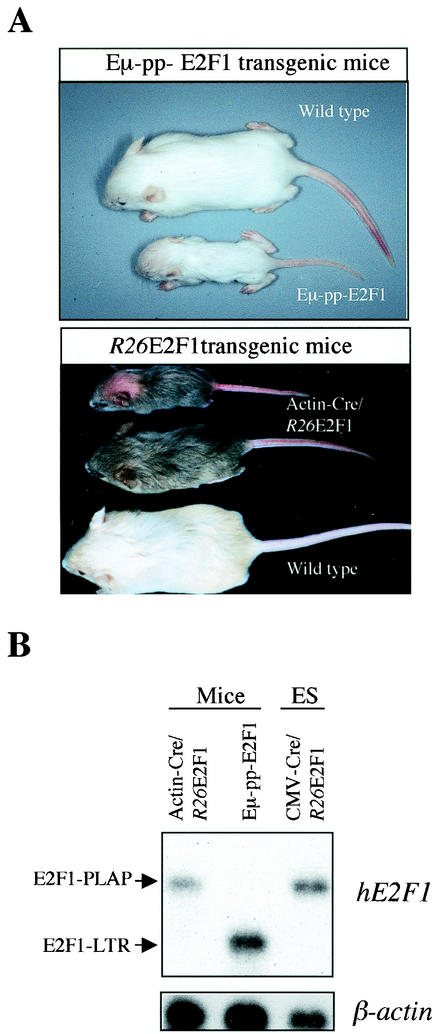
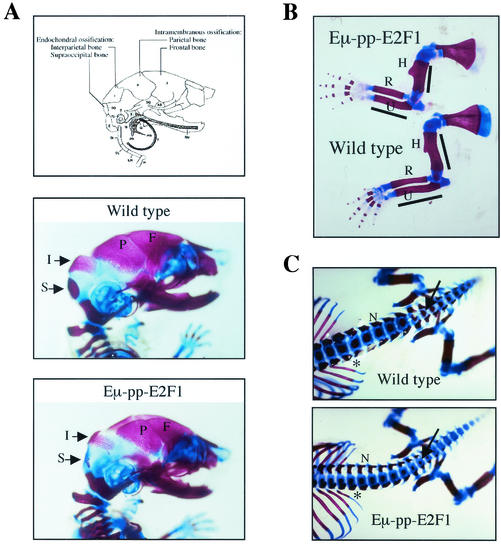
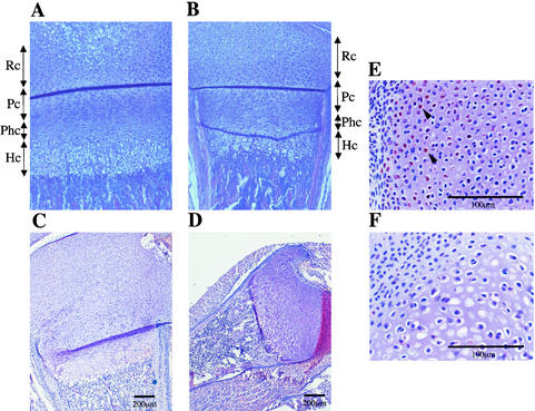
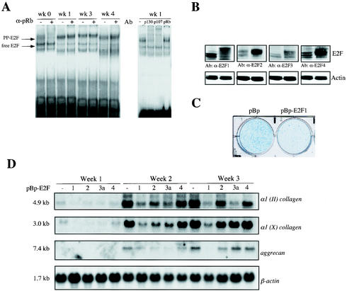
Longitudinal bone growth results from endochondral ossification, a process that requires proliferation and differentiation of chondrocytes. It has been shown that proper endochondral bone formation is critically dependent on the retinoblastoma family members p107 and p130. However, the precise functional roles played by individual E2F proteins remain poorly understood. Using both constitutive and conditional E2F1 transgenic mice, we show that ubiquitous transgene-driven expression of E2F1 during embryonic development results in a dwarf phenotype and significantly reduced postnatal viability. Overexpression of E2F1 disturbs chondrocyte maturation, resulting in delayed endochondral ossification, which is characterized by reduced hypertrophic zones and disorganized growth plates. Employing the chondrogenic cell line ATDC5, we investigated the effects of enforced E2F expression on the different phases of chondrocyte maturation that are normally required for endochondral ossification. Ectopic E2F1 expression strongly inhibits early- and late-phase differentiation of ATDC5 cells, accompanied by diminished cartilage nodule formation as well as decreased type II collagen, type X collagen, and aggrecan gene expression. In contrast, overexpression of E2F2 or E2F3a results in only a marginal delay of chondrocyte maturation, and increased E2F4 levels have no effect. These data are consistent with the notion that E2F1 is a regulator of chondrocyte differentiation.


Figures

FIG. 1.
FIG. 1.
Generation of Eμ-pp-E2F1 transgenic mice. (A) Transgene expression vector contains a duplicate version of the immunoglobulin heavy-chain enhancer (Eμ) inserted in the mouse pim1 promoter region (pp), followed by the Moloney murine leukemia virus long terminal repeat (LTR), which provides transcription termination sequences. The 3′ end of the human HA-E2F1 cDNA is fused to the SV40 small t antigen intervening sequence (IVS) of the pJ3Ω vector and provides an extra splice donor (SD) and splice acceptor (SA) site. (B) Immunoblotting shows E2F1 expression levels in total cell extracts from different tissues of 4-day-old Eμ-pp-E2F1 transgenic mice of two independent founder lines (TEF1-5 and TEF1-43) compared to control littermates. Actin serves as a loading control. Thym, thymus.
FIG. 2.
FIG. 2.
Conditional ROSA26-E2F1 transgenic mice. (A) (I) Structure of the ROSA26 (R26) locus, the R26-en2SA-loxP-E2F1-loxP-E2F1-PLAP targeting construct, and targeted locus. Restriction sites that have been inactivated are indicated with an asterisk. The position and orientation of the loxP sites are represented by triangles. The location of probe pHA607 (PstI-SalI fragment) and the BglII restriction sites used for genotyping the mice are depicted. (II and III) Schematic representation of the genomic structure and mRNA transcripts generated from the ROSA26 promoter before (II) and after (III) Cre-mediated excision. The HA-E2F1 cDNA is transcribed together with the PLAP cDNA, because of their linkage through the internal ribosomal entry site (IRES). (B) Southern blot analysis of BglII-digested DNA from ES cell clones containing one targeted R26-βgeo-E2F1 allele before and after Cre-mediated excision, compared to Cre-expressing control cells. Germ line (G), targeted (T), and excised (E) R26 alleles are indicated, as well as a nonspecific band recognized by probe pHA607 (asterisk). (C) Immunoblotting shows E2F1 expression levels in control, R26-βgeo-E2F1, and stable Cre-expressing R26E2F1 ES cells. Total cell lysate from CMV-HA-E2F1-transfected U2OS cells serves as a positive control. (D) Southern and Northern blot analyses on tissues isolated from Actin-Cre/R26E2F1;+ heterozygous, Actin-Cre/R26E2F1;R26E2F1 homozygous, and control (+/+) mice at the age of 5 weeks. The positions of the germ line (G) and excised (E) R26 alleles when probe pHA607 was used on BglII-digested genomic DNA are indicated to the left of the top blot. Northern blots demonstrate transgenic human E2F1 mRNA levels in several tissues, including spleen, liver, lung, midbrain, and bone marrow (BM), compared to β-actin loading control.
FIG. 3.
FIG. 3.
Phenotypes of E2F1 transgenic mice. (A) Gross physical appearance of 3-week-old Eμ-pp-E2F1 and Actin-Cre/R26E2F1/+ mice. Variability in the dwarf phenotype is demonstrated for the Actin-Cre/R26E2F1;+ mice. The bald spot on the smallest Actin-Cre/R26-E2F1;+ mouse was the result of excessive nurturing by the mother. (B) Northern blot analysis on poly(A)+ mRNA isolated from neck and trunk shows transgenic E2F1 gene expression in newborn Actin-Cre/R26E2F1;+ and Eμ-pp-E2F1 mice. RNA sample of CMV-Cre/R26E2F1;+ ES cells serves as a positive control for the 4.5-kb E2F1-IRES-PLAP transcript.
FIG. 4.
FIG. 4.
Skeletal analysis on E17.5 Eμ-pp-E2F1 transgenic mice. (A) Schematic representation of skull indicates distinct forms of ossification in interparietal (I) and supraoccipital (S) bones versus parietal (P) and frontal (F) bones. Lateral views of wild-type and Eμ-pp-E2F1 skulls stained with Alizarin red (calcified tissues) and Alcian blue (cartilage) are shown. (B) Lateral view of Eμ-pp-E2F1 and wild-type forelimbs. The humerus (H), radius (R), and ulna (U) are indicated. (C) Dorsoventral view of lumbosacral and tail vertebra showing reduced width of neural arches (N) as well as diminished degrees of ossification in distal vertebral bodies (arrow) and rib shaft regions (asterisk) of E2F1 transgenic mice compared to that of wild-type control mice.
FIG. 5.
FIG. 5.
Histological analysis of epiphyseal growth plates of E2F1 transgenic mice. (A and B) Sections of the growth plates of the femurs of 7-day-old wild-type mice (A) and Actin-Cre/R26-E2F1;+ mice (B) were stained with hematoxylin and eosin (H&E). Chondrocytes can be divided in four distinct zones: resting (Rc), proliferating (Pc), maturing prehypertrophic (Phc), and hypertrophic (Hc). The growth plates of Actin-Cre/R26-E2F1;+ mice display a relative larger zone of columnar proliferating chondrocytes and a shorter zone of prehypertrophic and hypertrophic chondrocytes. (C and D) H&E-stained sections of epiphyseal growth plates of wild-type (C) and Eμ-pp-E2F1 (D) newborn mice. Note the disorganization of hypertrophic zones within the smaller epiphysis of E2F1 transgenic mice. (E and F) Immunostaining with HA antibody on section of epiphysis of newborn Eμ-pp-E2F1 (E) and control wild-type (F) mice. Arrowheads indicate positively stained nuclei for HA-E2F1.
FIG. 6.
FIG. 6.
Ectopic expression of E2F transcription factors alters differentiation of chrondogenic ATDC5 cells. (A) Electrophoretic mobility shift analysis on extracts from prechondrogenic cells (week [wk] 0) and early (week 1) and late differentiating chondrocytes (week 3 and 4). The positions of the free E2F-DNA and pocket protein (PP)-E2F-DNA complexes are indicated by the arrows to the left of the gel. Supershift analyses with specific antibodies (Ab) against pRb (α-pRb) (21C9), p107 (C-18), and p130 (C-20) indicate the presence of distinct pocket protein complexes. (B) Western blot analysis shows expression of different E2F species in mock-infected pBabepuro ATDC5 cells (left lane of each blot) or polyclonal populations of ATDC5 cells infected with pBp-HA-E2F1, pBp-HA-E2F2, pBp-HA-E2F3a, or pBp-HA-E2F4 (right lane of each blot). Antibodies (Ab) against E2F1 (α-E2F1), E2F2, E2F3a, and E2F4 were used. (C) Cultures of polyclonal cells infected with pBabepuro (pBp) and pBp-HA-E2F1 were stained with Alcian blue after differentiation for 15 days, which detects differentiated cartilage nodules. (D) Expression of cartilage markers at different phases of chondrocyte differentiation in ATDC5 cells, which stably overexpress pBabepuro (pBp), pBp-HA-E2F1, pBp-HA-E2F2, pBp-HA-E2F3a, and pBp-HA-E2F4 (−, 1, 2, 3a, and 4, respectively). Total RNA was isolated at the indicated times and subjected to Northern blot analysis. Filters were serially hybridized with cDNA probes for type II collagen [α1 (II) collagen]), type X collagen [α1 (X) collagen], and aggrecan genes and β-actin, which serves as a loading control. Transcript sizes are indicated to the left of the blots.

References

    1. Akiyama, H., M. C. Chaboissier, J. F. Martin, A. Schedl, and B. De Crombrugghe. 2002. The transcription factor Sox9 has essential roles in successive steps of the chondrocyte differentiation pathway and is required for expression of Sox5 and Sox6. Genes Dev. 16:2813-2828. - PMC - PubMed
    1. Alkema, M. J., N. M. van der Lugt, R. C. Bobeldijk, A. Berns, and M. van Lohuizen. 1995. Transformation of axial skeleton due to overexpression of bmi-1 in transgenic mice. Nature 374:724-727. - PubMed
    1. Alvarez, J., P. Sohn, X. Zeng, T. Doetschman, D. J. Robbins, and R. Serra. 2002. TGFbeta2 mediates the effects of hedgehog on hypertrophic differentiation and PTHrP expression. Development 129:1913-1924. - PubMed
    1. Amanullah, A., B. Hoffman, and D. A. Liebermann. 2000. Deregulated E2F-1 blocks terminal differentiation and loss of leukemogenicity of M1 myeloblastic leukemia cells without abrogating induction of p15(INK4B) and p16(INK4A). Blood 96:475-482. - PubMed
    1. Amizuka, N., H. Warshawsky, J. E. Henderson, D. Goltzman, and A. C. Karaplis. 1994. Parathyroid hormone-related peptide-depleted mice show abnormal epiphyseal cartilage development and altered endochondral bone formation. J. Cell Biol. 126:1611-1623. - PMC - PubMed

Publication types

MeSH terms

LinkOut - more resources