. 2003 May 13;100(10):5828-33.
doi: 10.1073/pnas.1037282100. Epub 2003 Apr 30.

Generation of neural crest-derived peripheral neurons and floor plate cells from mouse and primate embryonic stem cells

Affiliations

Generation of neural crest-derived peripheral neurons and floor plate cells from mouse and primate embryonic stem cells

Kenji Mizuseki et al. Proc Natl Acad Sci U S A. .

Abstract

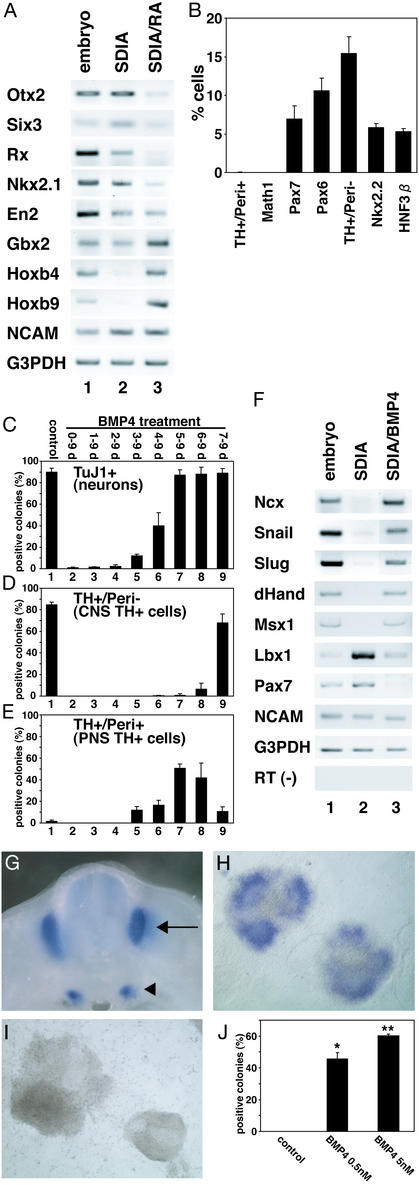
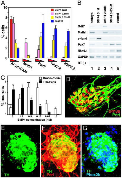
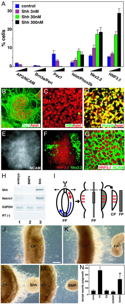
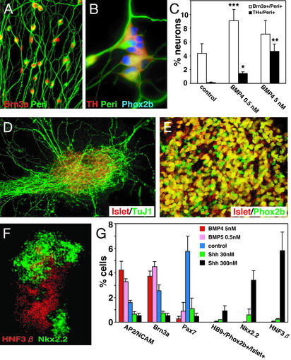
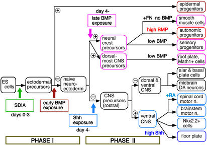
To understand the range of competence of embryonic stem (ES) cell-derived neural precursors, we have examined in vitro differentiation of mouse and primate ES cells into the dorsal- (neural crest) and ventralmost (floor plate) cells of the neural axis. Stromal cell-derived inducing activity (SDIA; accumulated on PA6 stromal cells) induces cocultured ES cells to differentiate into rostral CNS tissues containing both ventral and dorsal cells. Although early exposure of SDIA-treated ES cells to bone morphogenetic protein (BMP)4 suppresses neural differentiation and promotes epidermogenesis, late BMP4 exposure after the fourth day of coculture causes differentiation of neural crest cells and dorsalmost CNS cells, with autonomic system and sensory lineages induced preferentially by high and low BMP4 concentrations, respectively. In contrast, Sonic hedgehog (Shh) suppresses differentiation of neural crest lineages and promotes that of ventral CNS tissues such as motor neurons. Notably, high concentrations of Shh efficiently promote differentiation of HNF3beta(+) floor plate cells with axonal guidance activities. Thus, SDIA-treated ES cells generate naive precursors that have the competence of differentiating into the "full" dorsal-ventral range of neuroectodermal derivatives in response to patterning signals.


Figures

Figure 1
Figure 1
(A) RT-PCR analysis of SDIA-treated ES cells (day 8). Lane 1, embryonic day (E)10.5 embryo; lanes 2 and 3, SDIA-treated ES cells without and with 0.2 μM RA treatment during days 4–8, respectively. (B) Percent immunoreactive cells/total cells are shown. (C–E) BMP4 (0.5 nM) treatment during the days indicated above. Positive colonies were defined as ones containing TuJ1+ (C), TH+/Peri (D), and TH+/Peri+ (E) cells. Lane 1, control, no BMP4 treatment. (F) RT-PCR analysis with neural crest and neural markers. Lane 1, E10.5 whole embryos; lanes 2 and 3, SDIA-treated ES cells without or with 5 nM BMP4 treatment during days 5–8, respectively. (G) Ncx expression in E11.5 mouse by in situ hybridization. Dorsal root (arrow) and sympathetic (arrowhead) ganglia. (H) SDIA/BMP-treated ES cell colonies (day 8). (I) SDIA-treated ES cell colonies. (J) Percentages of Ncx+ colonies at 0, 0.5, and 5 nM BMP4 (n = 100, 200, and 200, respectively). *, P < 0.005 vs. control; **, P < 0.005 vs. control.
Figure 2
Figure 2
(A and C) Percent marker-positive cells in the colony (BMP4, days 5–9). Neurons were defined as TuJ1+ cells. *, P < 0.001 vs. 0 nM; **, P < 0.05 vs. 0.5 nM; ***, P < 0.001 vs. 0 nM; ****, P < 0.005 vs. 0.5 nM. (B) RT-PCR analysis of neural and neural crest markers. (D) Sensory lineage markers induced at a low BMP4 concentration [0.5 nM; immunostained with anti-Brn3a (red) and anti-Peri (green) antibodies]. (E–G) Cells with autonomic-lineage markers induced at a high BMP4 concentration (5 nM). Anti-TH (E–G, green); anti-Peri (F, red); anti-Phox2b (G, blue) antibodies.
Figure 3
Figure 3
(A) Effects of Shh (days 4–9) on neural crest and dorsal–ventral neural markers. (B–D) HB9/Phox2b+/Islet1/2+ motor neurons induced by Shh (30 nM, days 4–9). (E and F) Double staining of Nkx2.2 (green) and HNF3β (red) in an SDIA/Shh-treated ES cell colony. The NCAM staining in E indicates the colony area. (G) A high-magnification view. NCAM (green) and HNF3β (red). (H) RT-PCR analysis of Shh and Netrin expression. Lane 1, control embryo; lane 2, SDIA/BMP-treated; and lane 3, SDIA/Shh-treated ES cells. (I) A flat whole- mount preparation of embryonic rat brain and the explant coculture. Arrows, axonal direction. (J) Control CP explant; dorsal (right). (K) A floor plate explant showing chemoattraction activities on CP. (L) CP cocultured with SDIA/Shh-treated ES cells (Shh 300 nM). (M) CP cocultured with SDIA/BMP-treated ES cells (BMP4 5 nM). (N) Quantitative analysis of dorsally extending axon length from CP explants. Mean lengths of dorsal neurite outgrowth (micrometers) with CP alone (none, n = 12), FP (n = 12), undifferentiated ES (ES, n = 14), SDIA/BMP-treated ES (ES/BMP, n = 24), and SDIA/Shh-treated ES cells (ES/Shh, n = 28). *, P < 0.001 vs. none, ES, and ES/BMP.
Figure 4
Figure 4
(A–C) At low and high concentrations, BMP4 induced sensory bipolar neurons and autonomic neurons, respectively. Cells were treated with 0, 0.5, and 5 nM BMP4 during days 7–13 and analyzed on day 13 for Brn3a/Peri and TH/Peri staining, respectively. Neurons were defined as TuJ1+ cells. *, P < 0.001 vs. control; **, P < 0.005 vs. BMP4 0.5 nM; ***, P < 0.001 vs. control. (D and E) Islet1/2+/Phox2b+ motor neurons generated by SDIA/Shh treatment. (F) Double staining of Nkx2.2 (green) and HNF3β (red) in an SDIA/Shh-treated primate ES cell colony. (G) Effects of BMP4 and Shh on expression of dorsal and ventral markers. BMP4 and Shh were added during days 7–13 and 3–13, respectively.
Figure 5
Figure 5
Strategy for systematic induction of neural crest, dorsalmost CNS, and ventralmost CNS differentiation by timely exposure to patterning factors.

References

    1. Kawasaki H, Mizuseki K, Nishikawa S, Kaneko S, Kuwana Y, Nakanishi S, Nishikawa S-I, Sasai Y. Neuron. 2000;28:31–40. - PubMed
    1. Kawasaki H, Suemori H, Mizuseki K, Watanabe K, Urano F, Ichinose H, Haruta M, Takahashi M, Yoshikawa K, Nishikawa S-I, et al. Proc Natl Acad Sci USA. 2002;99:1580–1585. - PMC - PubMed
    1. Lee S-H, Lumelsky N, Studer L, Auerbach J M, McKay R D. Nat Biotechnol. 2000;18:675–679. - PubMed
    1. Wichterie H, Liberam I, Porter J A, Jessell T M. Cell. 2002;110:385–397. - PubMed
    1. Suemori H, Tada T, Torii R, Hosoi Y, Kobayashi K, Imahie H, Kondo Y, Iritani A, Nakatsuji N. Dev Dyn. 2001;222:273–279. - PubMed

Publication types