

Anthrax lethal factor represses glucocorticoid and progesterone receptor activity
- PMID: 12724519
- PMCID: PMC156265
- DOI: 10.1073/pnas.1036973100
Anthrax lethal factor represses glucocorticoid and progesterone receptor activity
Abstract
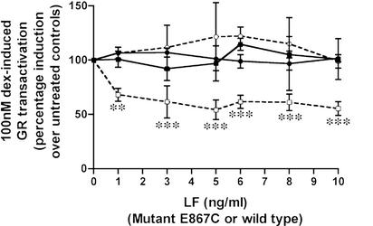
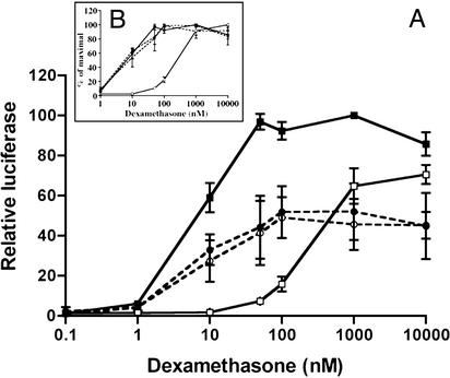
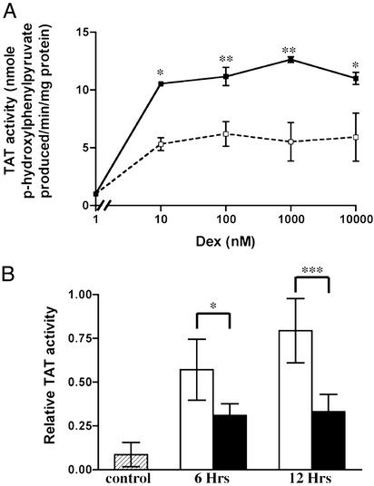
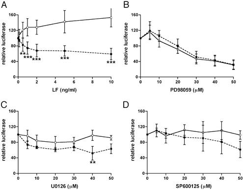
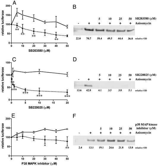
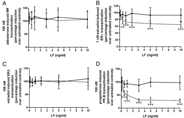
We report here that a bacterial toxin, anthrax lethal toxin (LeTx), at very low concentrations represses glucocorticoid receptor (GR) transactivation in a transient transfection system and the activity of an endogenous GR-regulated gene in both a cellular system and an animal model. This repression is noncompetitive and does not affect ligand binding or DNA binding, suggesting that anthrax lethal toxin (LeTx) probably exerts its effects through a cofactor(s) involved in the interaction between GR and the basal transcription machinery. LeTx-nuclear receptor repression is selective, repressing GR, progesterone receptor B (PR-B), and estrogen receptor alpha (ERalpha), but not the mineralocorticoid receptor (MR) or ERbeta. GR repression was also caused by selected p38 mitogen-activated protein (MAP) kinase inhibitors, suggesting that the LeTx action may result in part from its known inactivation of MAP kinases. Simultaneous loss of GR and other nuclear receptor activities could render an animal more susceptible to lethal or toxic effects of anthrax infection by removing the normally protective antiinflammatory effects of these hormones, similar to the increased mortality seen in animals exposed to both GR antagonists and infectious agents or bacterial products. These finding have implications for development of new treatments and prevention of the toxic effects of anthrax.
Figures
Similar articles
-
Novel repression of the glucocorticoid receptor by anthrax lethal toxin.Ann N Y Acad Sci. 2004 Jun;1024:9-23. doi: 10.1196/annals.1321.003. Ann N Y Acad Sci. 2004. PMID: 15265771 Review.
-
Anthrax lethal toxin represses glucocorticoid receptor (GR) transactivation by inhibiting GR-DNA binding in vivo.Mol Cell Endocrinol. 2005 Sep 28;241(1-2):21-31. doi: 10.1016/j.mce.2005.03.011. Mol Cell Endocrinol. 2005. PMID: 15964137
-
The large clostridial toxins from Clostridium sordellii and C. difficile repress glucocorticoid receptor activity.Infect Immun. 2007 Aug;75(8):3935-40. doi: 10.1128/IAI.00291-07. Epub 2007 May 21. Infect Immun. 2007. PMID: 17517870 Free PMC article.
-
Anthrax mounts a nuclear attack on glucocorticoid signaling.Trends Pharmacol Sci. 2003 Nov;24(11):558-9. doi: 10.1016/j.tips.2003.09.009. Trends Pharmacol Sci. 2003. PMID: 14607073 Review.
-
Anthrax lethal toxin disrupts the endothelial permeability barrier through blocking p38 signaling.J Cell Physiol. 2012 Apr;227(4):1438-45. doi: 10.1002/jcp.22859. J Cell Physiol. 2012. PMID: 21618534 Free PMC article.
Cited by
-
An overview of anthrax infection including the recently identified form of disease in injection drug users.Intensive Care Med. 2012 Jul;38(7):1092-104. doi: 10.1007/s00134-012-2541-0. Epub 2012 Apr 24. Intensive Care Med. 2012. PMID: 22527064 Free PMC article. Review.
-
Cellular and physiological effects of anthrax exotoxin and its relevance to disease.Front Cell Infect Microbiol. 2012 Jun 1;2:76. doi: 10.3389/fcimb.2012.00076. eCollection 2012. Front Cell Infect Microbiol. 2012. PMID: 22919667 Free PMC article. Review.
-
Anthrax lethal toxin induces endothelial barrier dysfunction.Am J Pathol. 2005 Jun;166(6):1871-81. doi: 10.1016/S0002-9440(10)62496-0. Am J Pathol. 2005. PMID: 15920171 Free PMC article.
-
Cellular and systemic effects of anthrax lethal toxin and edema toxin.Mol Aspects Med. 2009 Dec;30(6):439-55. doi: 10.1016/j.mam.2009.07.003. Epub 2009 Jul 26. Mol Aspects Med. 2009. PMID: 19638283 Free PMC article. Review.
-
Anthrax toxin receptor 2-dependent lethal toxin killing in vivo.PLoS Pathog. 2006 Oct;2(10):e111. doi: 10.1371/journal.ppat.0020111. PLoS Pathog. 2006. PMID: 17054395 Free PMC article.
References
MeSH terms
Substances
LinkOut - more resources
Full Text Sources
Other Literature Sources
Research Materials
