. 2003 May 13;100(10):6157-62.
doi: 10.1073/pnas.1031598100. Epub 2003 Apr 30.

Modulation of mitochondrial function by endogenous Zn2+ pools

Affiliations

Modulation of mitochondrial function by endogenous Zn2+ pools

Stefano L Sensi et al. Proc Natl Acad Sci U S A. .

Abstract

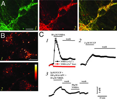
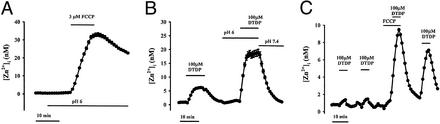
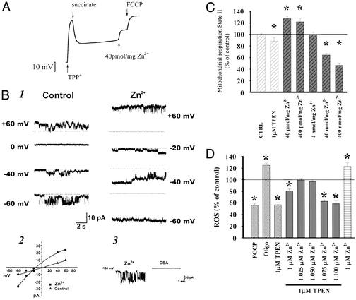
Recent evidence suggests that intracellular Zn(2+) accumulation contributes to the neuronal injury that occurs in epilepsy or ischemia in certain brain regions, including hippocampus, amygdala, and cortex. Although most attention has been given to the vesicular Zn(2+) that is released into the synaptic space and may gain entry to postsynaptic neurons, recent studies have highlighted pools of intracellular Zn(2+) that are mobilized in response to stimulation. One such Zn(2+) pool is likely bound to cytosolic proteins, like metallothioneins. Applying imaging techniques to cultured cortical neurons, this study provides novel evidence for the presence of a mitochondrial pool distinct from the cytosolic protein or ligand-bound pool. These pools can be pharmacologically mobilized largely independently of each other, with Zn(2+) release from one resulting in apparent net Zn(2+) transfer to the other. Further studies found evidence for complex and potent effects of Zn(2+) on isolated brain mitochondria. Submicromolar levels, comparable to those that might occur on strong mobilization of intracellular compartments, induced membrane depolarization (loss of Deltapsi(m)), increases in currents across the mitochondrial inner membrane as detected by direct patch clamp recording of mitoplasts, increased O(2) consumption and decreased reactive oxygen species (ROS) generation, whereas higher levels decreased O(2) consumption and increased ROS generation. Finally, strong mobilization of protein-bound Zn(2+) appeared to induce partial loss of Deltapsi(m), suggesting that movement of Zn(2+) between cytosolic and mitochondrial pools might be of functional significance in intact neurons.


Figures

Figure 1
Figure 1
Mobilization of Zn2+ from mitochondria. (A) Colocalization of RhodZin-3 and MitoTracker Green. Cultures were coloaded with the new Zn2+-sensitive mitochondrial probe RhodZin-3 (red fluorescence) and the mitochondrial marker, MitoTracker Green (green fluorescence), and imaged with confocal microscopy. Note the substantial overlap between these probes (yellow), indicating that they largely target the same intracellular organelles. Image is representative of 16 neurons from six experiments. (Bar = 10 μm.) (B) Mitochondria contain chelatable Zn2+. Cultures were loaded with RhodZin-3 and imaged under confocal microscopy before (1) and after (2) application of the cell-permeable Zn2+ chelator TPEN (20 μM). Note the decrement in speckled (mitochondrial) signal by TPEN, suggesting the presence of an intramitochondrial pool of chelatable Zn2+. Images are representative of 180 neurons from 11 experiments. (Bar = 10 μm; the pseudocolor bar shows the 12-bit fluorescence intensity range.) (C) Ca2+-dependent release of intramitochondrial Zn2+. FluoZin-3 loaded cultures were imaged before, during, and after 10-min exposures to NMDA (50 μM; 1), in a Ca2+-containing (black) or nominally Ca2+-free (red) buffer, to the mitochondrial protonophore, FCCP (3 μM; 2), or to FCCP followed by NMDA (3). Note that NMDA-induced Zn2+ rises depend on Ca2+ influx, and that prior exposure to FCCP occludes subsequent NMDA-triggered [Zn2+]i rises, indicating that these two manipulations target a common intracellular Zn2+ pool. Traces show time course of [Zn2+]i rises (±SEM) and are derived from 31–113 neurons from one experiment representative of 3–10.
Figure 2
Figure 2
Mobilization of mitochondrial and protein-bound Zn2+ pools. (A and B) Acidosis greatly enhances intracellular Zn2+ mobilization. (A) FluoZin-3-loaded cultures were exposed to FCCP (3 μM) after a 10-min preincubation in acidic (pH 6.0) buffer. Note that the [Zn2+]i rise is nearly an order of magnitude greater than that induced by an identical exposure at physiological pH (Fig. 1C; Table 1). (B) FluoZin-3-loaded cultures were exposed to the oxidizing agent, DTDP (10 min, 100 μM) at physiological pH, and after a 10-min preincubation at pH 6.0. Note the increased [Zn2+]i response at pH 6 and the rapid recovery on restoration of physiological pH. (C) Relocation of Zn2+ from mitochondria to protein-bound sites. FluoZin-3-loaded cultures were exposed to consecutive 5-min pulses of DTDP (100 μM), FCCP (3 μM), and both before a final exposure to DTDP, as indicated. Note that combined exposure to DTDP and FCCP induced far greater [Zn2+]i rises than exposure to FCCP alone, suggesting that in the absence of oxidation, Zn2+ released from mitochondria gets rapidly bound by redox-sensitive proteins. Further note the markedly increased size of the final DTDP response, indicative of a relocation of Zn2+ from the mitochondrial compartment to redox-sensitive protein pools. Traces show mean [Zn2+]i (±SEM) of 63–83 neurons, from 1 experiment representative of 5–14.
Figure 3
Figure 3
Potent effects of Zn2+ on isolated brain mitochondria. (A) Zn2+ depolarizes isolated brain mitochondria. Changes in Δψm are assessed by redistribution of TPP+ between the medium and the mitochondrial matrix. On addition of succinate as substrate, mitochondria generate Δψm (as indicated by the downward deflection of the trace). After a single pulse of Zn2+ (40 pmol/mg mitochondrial protein, likely corresponding to submicromolar free Zn2+) mitochondria undergo a sustained partial loss of Δψm, in comparison to the full loss of Δψm that occurs on addition of FCCP. Trace shows one experiment representative of six. (B) Zn2+ increases conductance and channel open probability of inner mitochondrial membranes. Traces show channel activity recorded in a patch of an isolated mitoplast from rat brain in the presence of 3 mM EGTA alone (Left) and in the presence of 300 nM Zn2+ (Right) (1). The patch was held at different voltages indicated (potentials refer to those of the patch electrode), and the dotted lines indicate the closed state. The membrane conductance and channel open probability are both increased. Current–voltage relationship for the two sets of recordings shown in 1 (2). CSA inhibits channel activity activated by Zn2+ (3). The traces show activity induced by 300 nM Zn2+ before (Left) and after (Right) addition of CSA (5 μM) to the bath. (C) Effects of Zn2+ on mitochondrial respiration. Isolated brain mitochondria are energized with 5 mM pyruvate/2.5 mM malate, and O2 consumption measured with an O2-sensitive electrode in the presence of TPEN or Zn2+ as indicated. Note the minimal decrease in respiratory rate induced by TPEN, as well as the biphasic effect of Zn2+, with low levels increasing and higher levels decreasing respiration. (D) Effects of Zn2+ on mitochondrial ROS generation. Isolated brain mitochondria are energized as above, and ROS generation is measured as changes in fluorescence of the oxidation-sensitive dye, H2DCFDA in the presence of FCCP (1 μM), oligomycin (1 μM), TPEN (1 μM), and/or Zn2+, as indicated. Note the decrease in ROS production induced by FCCP, consistent with uncoupling, and the expected increase in ROS generation caused by the mitochondrial ATPase inhibitor oligomycin (which hyperpolarizes mitochondria). Further note the decrease in ROS generation caused by TPEN, its reversal by addition of equimolar Zn2+, the decrease in ROS generation caused by a slight excess of the cation over TPEN (consistent with uncoupling), and finally the increase in ROS generation by addition of a larger amount of Zn2+ (consistent with inhibition of electron transport). In C and D, data were analyzed by using an ANOVA and subsequent Fisher's least significant difference test (n = 6). Bars represent group means ± SEM; asterisks indicate P < 0.01.
Figure 4
Figure 4
Mobilization of protein-bound Zn2+ triggers partial loss of Δψm. Cultures were loaded with the Δψm-sensitive probe rhodamine 123 (an increase in fluorescence indicates loss of Δψm), and exposed to DTDP (100 μM) in an acidic environment (pH 6.0) to produce a strong mobilization of protein- bound Zn2+ pools. DTDP-induced rhodamine 123 fluorescence changes were compared with those observed on full loss of Δψm elicited by addition of FCCP (3 μM) at the end of the experiment. Note that inclusion of TPEN (20 μM) during the exposure substantially attenuated the rhodamine 123 fluorescence increase (Upper). Traces show mean (±SEM) of rhodamine 123 fluorescence changes from of 58–64 neurons from one experiment representative of 11. Bar graph (Lower) shows compiled rhodamine 123 fluorescence changes (means ± SEM), expressed as percent of maximal change induced by FCCP, at the end of 20-min DTDP exposures as above, at pH 7.4 and pH 6 (n = 567–964 in 8–11 experiments, asterisk indicates P < 0.01 by ANOVA and subsequent Bonferroni's test).

References

    1. Frederickson C J, Bush A I. Biometals. 2001;14:353–366. - PubMed
    1. Lee J Y, Cole T B, Palmiter R D, Koh J Y. J Neurosci. 2000;20:RC79. - PMC - PubMed
    1. Aschner M, Cherian M G, Klaassen C D, Palmiter R D, Erickson J C, Bush A I. Toxicol Appl Pharmacol. 1997;142:229–242. - PubMed
    1. Coyle P, Philcox J C, Carey L C, Rofe A M. Cell Mol Life Sci. 2002;59:627–647. - PMC - PubMed
    1. Kagi J H R. In: Metallothionein III: Biological Roles and Medical Implications. Kimura M, editor. Basel: Birkhauser; 1993. pp. 29–55.

Publication types

LinkOut - more resources