. 2003 May 13;100(10):6121-6.
doi: 10.1073/pnas.1030024100. Epub 2003 Apr 30.

MmpL8 is required for sulfolipid-1 biosynthesis and Mycobacterium tuberculosis virulence

Affiliations

MmpL8 is required for sulfolipid-1 biosynthesis and Mycobacterium tuberculosis virulence

Scott E Converse et al. Proc Natl Acad Sci U S A. .

Abstract

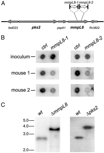
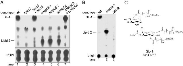
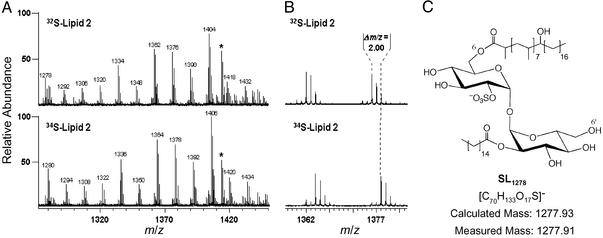
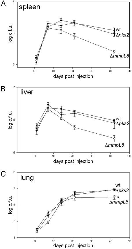
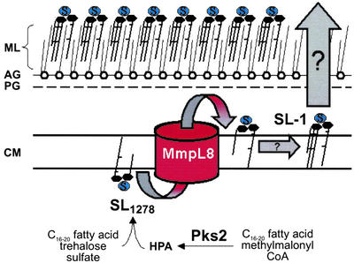
Mycobacterium tuberculosis, the causative agent of human tuberculosis, is unique among bacterial pathogens in that it displays a wide array of complex lipids and lipoglycans on its cell surface. One of the more remarkable lipids is a sulfated glycolipid, termed sulfolipid-1 (SL-1), which is thought to mediate specific host-pathogen interactions during infection. However, a direct role for SL-1 in M. tuberculosis virulence has not been established. Here we show that MmpL8, a member of a large family of predicted lipid transporters in M. tuberculosis, is required for SL-1 production. The accumulation of an SL-1 precursor, termed SL(1278), in mmpL8 mutant cells indicates that MmpL8 is necessary for an intermediate step in the SL-1 biosynthesis pathway. We use a novel fractionation procedure to demonstrate that SL-1 is present on the cell surface, whereas SL(1278) is found exclusively in more internal layers. Importantly, we show that mmpL8 mutants are attenuated for growth in a mouse model of tuberculosis. However, SL-1 per se is not required for establishing infection as pks2 mutants, which are defective in an earlier step in SL-1 biosynthesis, have no obvious growth defect. Thus, we hypothesize that either MmpL8 transports molecules in addition to SL-1 that mediate host-pathogen interactions or the accumulation of SL(1278) in mmpL8 mutant cells interferes with other pathways required for growth during the early stages of infection.


Figures

Figure 1
Figure 1
Two mmpL8 mutants of M. tuberculosis identified by signature-tagged mutagenesis. (A) Diagram of mmpL8 and surrounding genes denoting the transposon insertion sites (1,660 and 2,238 bp with respect to the start codon) in each mutant. (B) Signature DNA tags from mutant mycobacteria harvested from the inoculum and the lungs of two mice after 3 weeks of growth were amplified, radiolabelled, and hybridized to tag array filters (18). Tags from the two underrepresented mmpL8 mutants are shown next to control tags that did not vary throughout the experiment. (C) ΔmmpL8 and Δpks2 strains were generated by specialized transduction, and Southern blot revealed the expected bands indicating the replacement of the genomic sequence with the hygromycin cassette. For details, see Materials and Methods.
Figure 2
Figure 2
TLC analysis of lipids from mmpL8 and pks2 M. tuberculosis mutants is shown. (A) Cells were labeled with 14C-propionate, and lipids were extracted and separated under two TLC solvent conditions. The first (Upper) resolves SL-1, whereas the second (Lower) resolves PDIM. Equal amounts of lipids were spotted onto both TLCs. The position of SL-1, lipid 2, and the two forms of PDIM are noted. (B) Cells were labeled with 35S-SOformula image, and lipids were extracted and separated by TLC, as in A Upper. (C) Proposed structure of SL-1 as determined by Mougous et al. (23) and Goren et al. (25, 26).
Figure 3
Figure 3
Structural characterization of lipid 2 by MS is shown. (A) Cells were labeled with 32S-SOformula image or 34S-SOformula image, lipid 2 was partially purified by preparative TLC, and both lipid samples were analyzed by Fourier transform ion cyclotron resonance MS. Comparison of both spectra revealed a single group of species (m/z values denoted above each peak) that shifted by 2 mass units, indicating the presence of one sulfur atom in each molecule. An unrelated, sulfur-free compound within the spectra is marked with an asterisk. (B) A magnified view of the spectra from A that shows two peaks that shifted when cells were labeled with 34S-SOformula image. (C) Proposed structure of the SL-related lipid 2 species, SL1278, as determined by exact mass analysis. The mass measured by MS differs from the calculated mass of the proposed structure by 11 ppm.
Figure 4
Figure 4
SL1278 is a biosynthetic precursor of SL-1 and is not exposed on the mycobacterial cell surface. (A) Wild-type (Upper) and ΔmmpL8 mutant (Lower) cells were pulsed labeled with 14C-propionate for 30 min, washed to remove unincorporated label, and grown in liquid media. At the times indicated during the chase, cells were harvested, and lipids were extracted and analyzed by TLC. The ΔmmpL8 sample at t = 60 is underloaded, not deficient in SL1278. (B) Cells were labeled with 14C-propionate for 2 h, and surface-exposed lipids (S) were extracted by using hexanes/decylamine and gentle sonication. Cell pellets (P) containing the lipids not extracted by this procedure were harvested by centrifugation and lipids from both fractions were extracted and analyzed by TLC.
Figure 5
Figure 5
mmpL8, but not pks2, is required for growth in a mouse model of tuberculosis. C57/B6 mice were infected by tail-vein injection with 1–2 × 10⁶ cells of wild-type (filled circles), ΔmmpL8 (open circles), or Δpks2 (filled triangles) mutant M. tuberculosis cells. Growth in the spleen (A), liver (B), and lung (C) was monitored by harvesting organs at 1, 7, 14, 21, and 42 days after infection, and bacillary loads were determined by plating dilutions on solid media. Each data point represents the average of colony-forming units from five infected mice, and error bars indicate the standard error of the means. The data shown here is from one experiment and a duplicative experiment gave nearly identical results. *, P value of 0.003.
Figure 6
Figure 6
Model of the role of MmpL8 in the SL-1 biosynthetic pathway. See Discussion for details of the model. HPA, hydroxyphthioceranic acid; ML, mycolate layer; AG, arabinogalactan layer; PG, peptidoglycan layer; CM, cytoplasmic membrane.

References

    1. Russell D G. Nat Rev Mol Cell Biol. 2001;2:569–577. - PubMed
    1. McKinney J D, Jacobs J, W R, Bloom B R. In: Emerging Infections. Fauci A, Krause R, editors. London: Academic; 1998. pp. 51–146.
    1. Glickman M S, Jacobs W R., Jr Cell. 2001;104:477–485. - PubMed
    1. Brennan P J, Nikaido H. Annu Rev Biochem. 1995;64:29–63. - PubMed
    1. Nikaido H, Kim S H, Rosenberg E Y. Mol Microbiol. 1993;8:1025–1030. - PubMed

Publication types

LinkOut - more resources