. 2003 May;15(5):1095-110.
doi: 10.1105/tpc.010413.

Dominant-negative receptor uncovers redundancy in the Arabidopsis ERECTA Leucine-rich repeat receptor-like kinase signaling pathway that regulates organ shape

Affiliations

Dominant-negative receptor uncovers redundancy in the Arabidopsis ERECTA Leucine-rich repeat receptor-like kinase signaling pathway that regulates organ shape

Elena D Shpak et al. Plant Cell. 2003 May.

Abstract

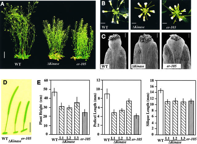
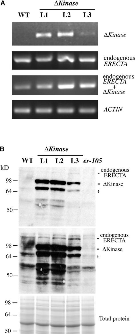
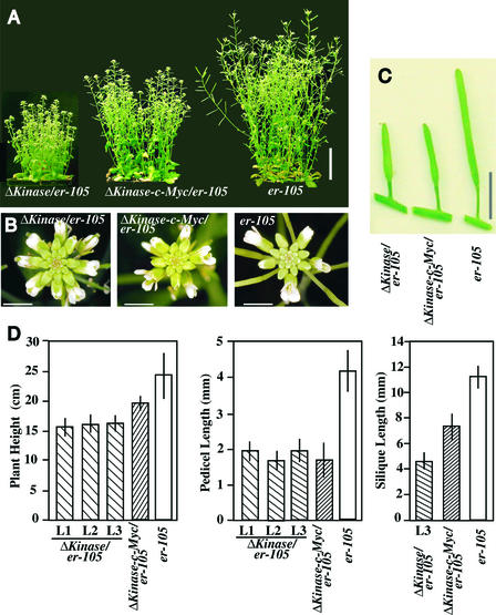
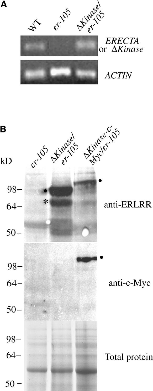
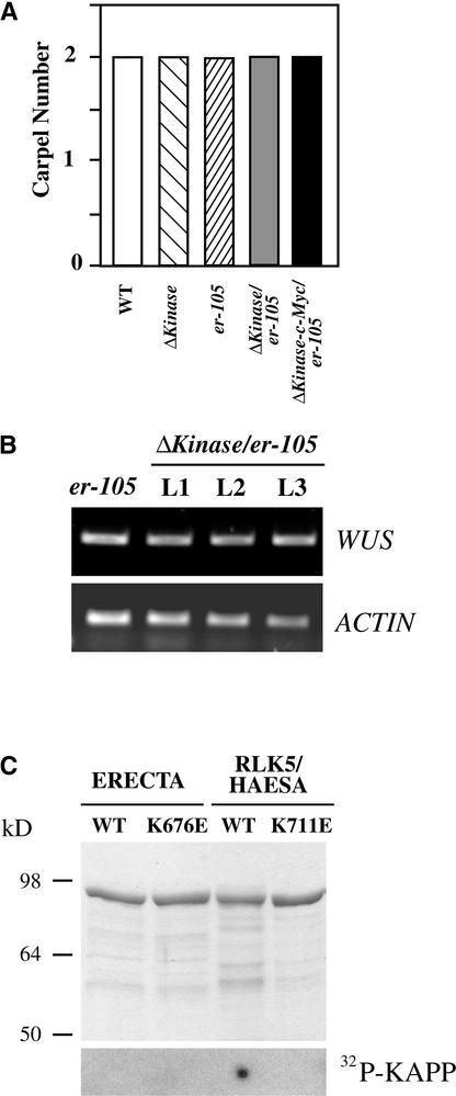
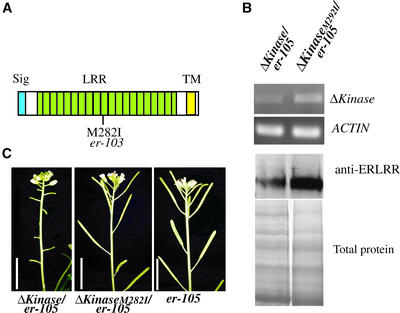
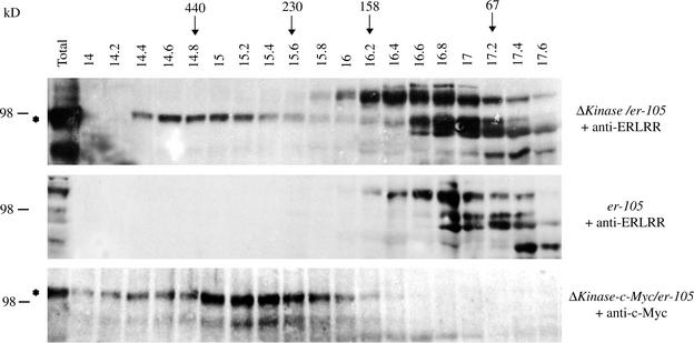
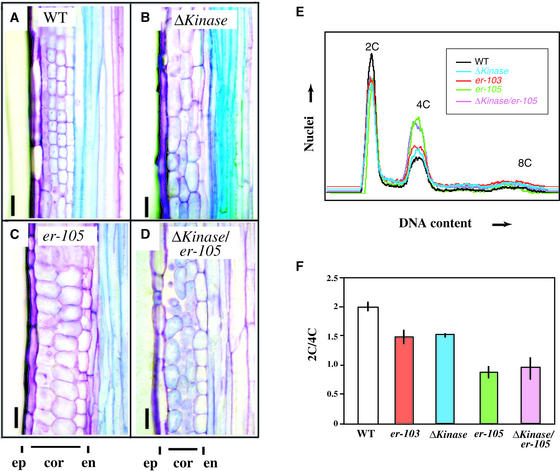
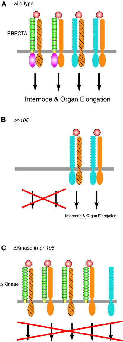
Arabidopsis ERECTA, a Leu-rich repeat receptor-like Ser/Thr kinase (LRR-RLK), regulates organ shape and inflorescence architecture. Here, we show that a truncated ERECTA protein that lacks the cytoplasmic kinase domain (DeltaKinase) confers dominant-negative effects when expressed under the control of the native ERECTA promoter and terminator. Transgenic plants expressing DeltaKinase displayed phenotypes, including compact inflorescence and short, blunt siliques, that are characteristic of loss-of-function erecta mutant plants. The DeltaKinase fragment migrated as a stable approximately 400-kD protein complex in the complete absence of the endogenous ERECTA protein and significantly exaggerated the growth defects of the null erecta plants. A functional LRR domain of DeltaKinase was required for dominant-negative effects. Accumulation of DeltaKinase did not interfere with another LRR-RLK signaling pathway (CLAVATA1), which operates in the same cells as ERECTA but has a distinct biological function. Both the erecta mutation and DeltaKinase expression conferred a lesser number of large, disorganized, and expanded cortex cells, which are associated with an increased level of somatic endoploidy. These findings suggest that functionally redundant RLK signaling pathways, including ERECTA, are required to fine-tune the proliferation and growth of cells in the same tissue type during Arabidopsis organogenesis.


Figures

Figure 1.
Figure 1.
ERECTA::ΔKinase Confers Dominant-Negative Effects in Arabidopsis Morphology. (A) Seven-week-old plants of the wild type (WT), ΔKinase, and erecta-105. Bar = 4 cm. (B) Inflorescence apices from the wild type, ΔKinase, and erecta-103. Bars = 3 mm. (C) Scanning electron micrographs of silique tips from the wild type, ΔKinase, and erecta-105. Bars = 100 μm. (D) Siliques and attached pedicels of the wild type, ΔKinase, and erecta-105. Bar = 5 mm. (E) Morphometric analysis of fully grown 7-week-old plants of the wild type, erecta-105, and three independent transgenic lines (L1, L2, and L3) of ERECTA::ΔKinase. Twenty-five plants were analyzed for plant (inflorescence) height. Lengths of 50 mature pedicels and siliques on the main inflorescence stem (10 measurements per stem) were analyzed. Bars represent average values ± sd.
Figure 2.
Figure 2.
Dominant-Negative Effects in ΔKinase Are Caused by the Presence of the Truncated Receptor and Not by the Cosuppression of Endogenous ERECTA. (A) Semiquantitative RT-PCR analysis of the ΔKinase and ERECTA transcripts in buds and flower clusters of the wild type (WT) and three independent ERECTA::ΔKinase lines (L1, L2, and L3). The top gel shows that expression levels of ΔKinase correlate with the severity of the phenotype. The second gel shows the lack of cosuppression revealed by primers that specifically amplify the endogenous ERECTA transcripts. In the third gel, primers that amplify both endogenous ERECTA and the ΔKinase transcripts revealed that ΔKinase transcripts are present at a level comparable to that in the endogenous ERECTA. The bottom gel represents an actin fragment amplified simultaneously as a control. (B) Immunoblot analysis of protein extracts from buds and flower clusters of the wild type, three independent ERECTA::ΔKinase lines, and the null allele erecta-105. In the top gel, a blot exposed for a short period (3 to 5 min) shows that the amount of the ΔKinase protein correlates with the amount of its message. The identical blot in the middle was exposed for a longer period (∼50 min) to detect the endogenous ERECTA protein, which was present at much lower levels compared with ΔKinase. At bottom, a Coomassie blue–stained gel shows the total proteins. Asterisks indicate cleaved ΔKinase protein. Numbers at left indicate molecular mass markers in kilodaltons.
Figure 3.
Figure 3.
Expression of ΔKinase Enhances the Growth Defects in the Null erecta Allele. (A) Seven-week-old plants of erecta-105, ΔKinase/erecta-105, and ΔKinase–c-Myc/erecta-105. Bar = 4 cm. (B) Inflorescence apices from erecta-105, ΔKinase/erecta-105, and ΔKinase–c-Myc/erecta-105. Bars = 3 mm. (C) Siliques and attached pedicels of erecta-105, ΔKinase/erecta-105, and ΔKinase–c-Myc/erecta-105. Bar = 5 mm. (D) Morphometric analysis of fully grown 7-week-old plants of erecta-105, three independent transgenic lines of ERECTA::ΔKinase/erecta-105 (L1, L2, and L3), and one line of ERECTA::ΔKinase–c-Myc/erecta-105. Plant (inflorescence) height, pedicel length, and silique length were measured as described for Figure 1E. The length of the siliques was measured in only one line of ERECTA::ΔKinase/erecta-105 (L3), because the other two lines had reduced fertility as a result of short filaments. Bars represent average values ± sd.
Figure 4.
Figure 4.
Accumulation of ΔKinase Transcripts and ΔKinase and ΔKinase–c-Myc Proteins in Transgenic erecta-105 Plants. (A) Semiquantitative RT-PCR analysis of ERECTA and ΔKinase transcripts from buds and flower clusters of wild-type (WT), erecta-105, and ERECTA::ΔKinase/erecta-105 plants. ΔKinase transcripts are expressed in the transgenic erecta-105 plants at a level similar to that of the endogenous ERECTA transcripts in wild-type plants. The erecta-105 plants do not express any ERECTA transcripts. Primers annealing to the LRR-coding region of ERECTA were used for PCR. An actin fragment was amplified simultaneously as a control. (B) Protein immunoblot analysis. At top, a blot probed with anti-ERLRR antibodies shows the accumulation of the ΔKinase and ΔKinase–c-Myc proteins in transgenic erecta-105 plants. The identical blot in the middle was probed with anti-c-Myc antibodies and shows the accumulation of the ΔKinase–c-Myc protein. At bottom, a Coomassie blue–stained gel shows the total proteins. Numbers at left indicate molecular mass markers in kilodaltons. Dots indicate the transgene products. An asterisk indicates cleaved ΔKinase protein.
Figure 5.
Figure 5.
Expression of ΔKinase Does Not Interfere with the CLV Signaling Pathway, and ERECTA Does Not Associate with KAPP, a Negative Regulator of Multiple RLKs, Including CLV1. (A) Number of carpels in wild-type (WT), erecta-105, ERECTA::ΔKinase/wild type (L1), ERECTA::ΔKinase/erecta-105 (L3), and ERECTA::ΔKinase–c-Myc/erecta-105 flowers. Bars represent mean values (n = 40) for each genotype. (B) Semiquantitative RT-PCR analysis of WUS transcripts from buds and flower clusters of erecta-105 and three independent lines of ERECTA::ΔKinase/erecta-105 plants. WUS expression levels are unaltered by the ΔKinase fragment. (C) KAPP KID does not associate with an active kinase catalytic domain of ERECTA. Top, Coomassie blue–stained SDS-PAGE gel of the affinity-purified, recombinant ERECTA and RLK5/HAESA kinase domains, both active (WT) and inactive (for ERECTA, K676E; for RLK5, K711E) forms, fused to the maltose binding protein. One microgram of the purified proteins was loaded on the gel. Molecular mass markers are indicated at left. Bottom, Autoradiogram of the purified proteins dot-blotted onto a polyvinylidene difluoride membrane and probed with the 32P-labeled GST-KID fusion protein.
Figure 6.
Figure 6.
A Functional LRR Domain of ΔKinase Is Required for the Dominant-Negative Interference. (A) Scheme showing the location of an introduced point mutation corresponding to erecta-103, which replaces the Met at amino acid 282 with Ile. TM, transmembrane domain; Sig, signal peptide. (B) The M282I mutation in ΔKinase does not lead to reduced transcript and protein levels. The top two gels show semiquantitative RT-PCR analysis of ΔKinase and control actin transcripts in buds and flower clusters of ERECTA::ΔKinase/erecta-105 (L1) and ERECTA::ΔKinaseM282I /erecta-105 plants. The third gel shows protein immunoblot analysis of extracts from buds and flower clusters of ERECTA::ΔKinase/erecta-105 (L1) and ERECTA::ΔKinaseM282I /erecta-105 plants probed with anti-ERLRR antibody. The Coomassie blue–stained gel at bottom shows the total proteins. (C) The M282I mutation in ΔKinase severely compromises the dominant-negative effects. Shown are side views of the main inflorescences of ERECTA::ΔKinase/erecta-105 (L1), ERECTA::ΔKinaseM282I /erecta-105, and erecta-105 plants. Bars = 1 cm.
Figure 7.
Figure 7.
Both ΔKinase and ΔKinase–c-Myc Proteins Migrate as an ∼400-kD Protein Complex in the Absence of the Endogenous ERECTA Protein. Total proteins (for ΔKinase) or membrane proteins (for ΔKinase–c-Myc) isolated from buds and flower clusters were separated by gel-filtration chromatography (see Methods). Each fraction was separated by SDS-PAGE. Total proteins were loaded in the first lane for comparison. Numbers above each lane refer to the elution volume (in milliliters) of the corresponding fraction. The elution peaks of molecular mass standards for gel-filtration chromatography are indicated at top (arrows). Molecular mass standards for SDS-PAGE are indicated at left. The top gel shows the detection of the ΔKinase complex (asterisk) in transgenic ERECTA::ΔKinase/erecta-105 plants on an immunoblot probed with anti-ERLRR antibodies. The middle gel shows a control immunoblot of erecta-105 fractions probed with anti-ERLRR antibodies. The bottom gel shows the detection of the ΔKinase–c-Myc complex (asterisk) in transgenic ERECTA::ΔKinasec-Myc/erecta-105 plants on an immunoblot probed with anti-c-Myc antibodies.
Figure 8.
Figure 8.
erecta Mutation and ΔKinase Expression Lead to Aberrant Cell Enlargement, Reduced Cell Numbers in the Cortex, and Increased Endoploidy in Mature Pedicels. (A) to (D) Longitudinal sections of mature pedicels from wild-type (WT) (A), ERECTA::ΔKinase (B), erecta-105 (C), and ERECTA::ΔKinase/erecta-105 (D) plants. Photographs were taken at the same magnification. cor, cortex; en, endodermis; ep, epidermis. Bars = 25 μm. (E) and (F) Flow cytometric analysis of mature pedicels from wild-type, intermediate allele erecta-103, ERECTA::ΔKinase, null allele erecta-105, and ERECTA::ΔKinase/erecta-105 plants. (E) shows representative nuclei counts, and (F) shows the 2C/4C ratio as an average of four independent counts. Error bars indicate standard errors.
Figure 9.
Figure 9.
Hypothetical Model for the ERECTA Signaling Pathway and ΔKinase Action. (A) In wild-type plants, ERECTA associates with multiple partners (orange). Additional RLKs (blue) may share ligands (red) as well as receptor partners of ERECTA. The activation of these pathways promotes internode/organ elongation. (B) erecta-105 plants do not have any ERECT. However, the receptor complex with overlapping function partially promotes internode/organ elongation. (C) In ERECTA::ΔKinase/erecta-105 plants, highly stable ERECTA ΔKinase fragments shut down the entire pathway, presumably by forming inactive receptor heterodimers and/or depleting ligands for the RLK that has a function partially redundant with that of ERECTA.

References

    1. Amaya, E., Musci, T., and Kirschner, M. (1991). Expression of a dominant-negative mutant of the FGF receptor disrupts mesoderm formation in Xenopus embryos. Cell 66, 257–270. - PubMed
    1. Ataliotis, P., and Mercola, M. (1997). Distribution and function of platelet-derived growth factors and their receptors during embryogenesis. Int. Rev. Cytol. 172, 95–125. - PubMed
    1. Azpiroz, R., Wu, Y., LoCascio, J.C., and Feldmann, K.A. (1998). An Arabidopsis brassinosteroid-dependent mutant is blocked in cell elongation. Plant Cell 10, 219–230. - PMC - PubMed
    1. Bechtold, N., Ellis, J.G., and Pelletier, G. (1993). In planta Agrobacterium-mediated gene transfer by infiltration of adult Arabidopsis thaliana plants. C. R. Acad. Sci. Paris 316, 1194–1199.
    1. Beguinot, L., Lyall, R., Willingham, M., and Pastan, I. (1984). Down-regulation of the epidermal growth factor receptor in KB cells is due to receptor internalization and subsequent degradation in lysosomes. Proc. Natl. Acad. Sci. USA 81, 2384–2388. - PMC - PubMed

Publication types

MeSH terms

LinkOut - more resources