

doi: 10.1016/s0168-1702(03)00065-0.
Sequence analysis of the N, P, M and F genes of Canadian human metapneumovirus strains
Affiliations
- PMID: 12727342
- PMCID: PMC7172423
- DOI: 10.1016/s0168-1702(03)00065-0
Item in Clipboard
Sequence analysis of the N, P, M and F genes of Canadian human metapneumovirus strains
Virus Res.
2003 May.
Abstract
The complete nucleotide sequences of the nucleoprotein (N), phosphoprotein (P), matrix protein (M), and fusion protein (F) genes of 15 Canadian human metapneumovirus (hMPV) isolates were determined. Phylogenetic analysis revealed two distinct genetic clusters, or groups for each gene with additional sequence variability within the individual groups. Comparison of the deduced amino acid sequences for the N, M and F genes of the different isolates revealed that all three genes were well conserved with 94.1-97.6% identity between the two distinct clusters The P gene showed more diversity with 81.6-85.7% amino acid identity for isolates between the two clusters, and 94.6-100% for isolates within the same cluster.
Figures
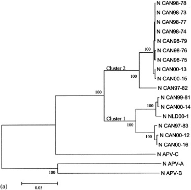
Phylogenic analysis of hMPV Canadian isolates. The N (a), P (b), M (c) and F (d) ORFs were amplified by PCR and sequenced. The corresponding gene sequences from APV subgroups and hMPV from the Netherlands (NLD00-1) were also analyzed. Bootstrap proportions were plotted at the main internal branches of the phylogram to show support values. Phylogenetic analysis was performed using the Neighbor-Joining method of the MEGA program. The year in which the isolate was collected is indicated by the first two numbers in the isolate name.
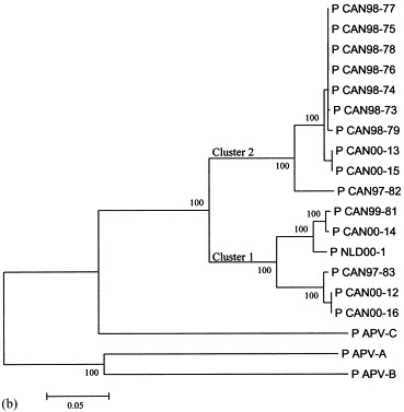
Phylogenic analysis of hMPV Canadian isolates. The N (a), P (b), M (c) and F (d) ORFs were amplified by PCR and sequenced. The corresponding gene sequences from APV subgroups and hMPV from the Netherlands (NLD00-1) were also analyzed. Bootstrap proportions were plotted at the main internal branches of the phylogram to show support values. Phylogenetic analysis was performed using the Neighbor-Joining method of the MEGA program. The year in which the isolate was collected is indicated by the first two numbers in the isolate name.
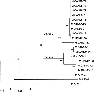
Phylogenic analysis of hMPV Canadian isolates. The N (a), P (b), M (c) and F (d) ORFs were amplified by PCR and sequenced. The corresponding gene sequences from APV subgroups and hMPV from the Netherlands (NLD00-1) were also analyzed. Bootstrap proportions were plotted at the main internal branches of the phylogram to show support values. Phylogenetic analysis was performed using the Neighbor-Joining method of the MEGA program. The year in which the isolate was collected is indicated by the first two numbers in the isolate name.
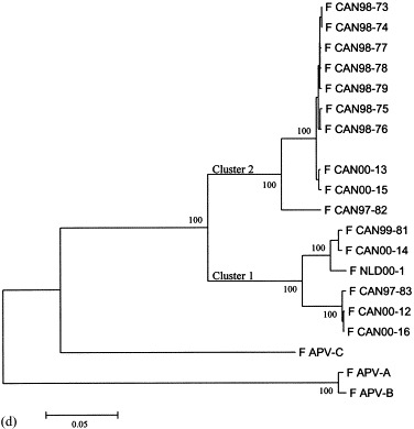
Phylogenic analysis of hMPV Canadian isolates. The N (a), P (b), M (c) and F (d) ORFs were amplified by PCR and sequenced. The corresponding gene sequences from APV subgroups and hMPV from the Netherlands (NLD00-1) were also analyzed. Bootstrap proportions were plotted at the main internal branches of the phylogram to show support values. Phylogenetic analysis was performed using the Neighbor-Joining method of the MEGA program. The year in which the isolate was collected is indicated by the first two numbers in the isolate name.
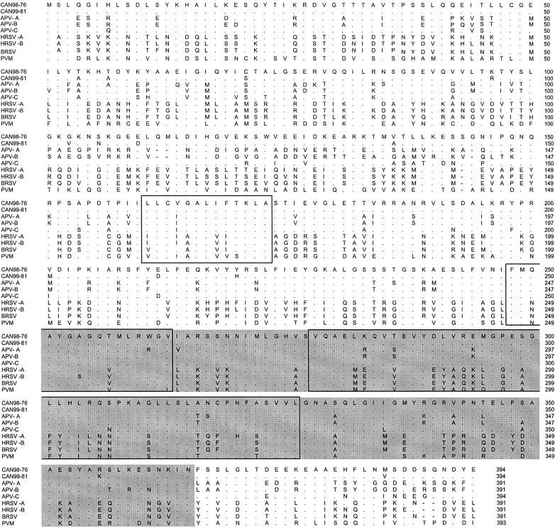
Alignment of the predicted amino acid sequences of the hMPV N protein of hMPV CAN99-81 (cluster 1) with those of CAN98-76 (cluster 2), APV-A, B and C, HRSV A and B, BRSV and PVM. Only residues that differ from isolate CAN98-76 are shown, identical amino acids are represented by periods, gaps are represented by dashes. A relatively conserved domain is shaded (residues 247–365). Three conserved domains present in all non-segmented, negative stranded viruses at residues 161–173, 251–263 and 279–326 are boxed.
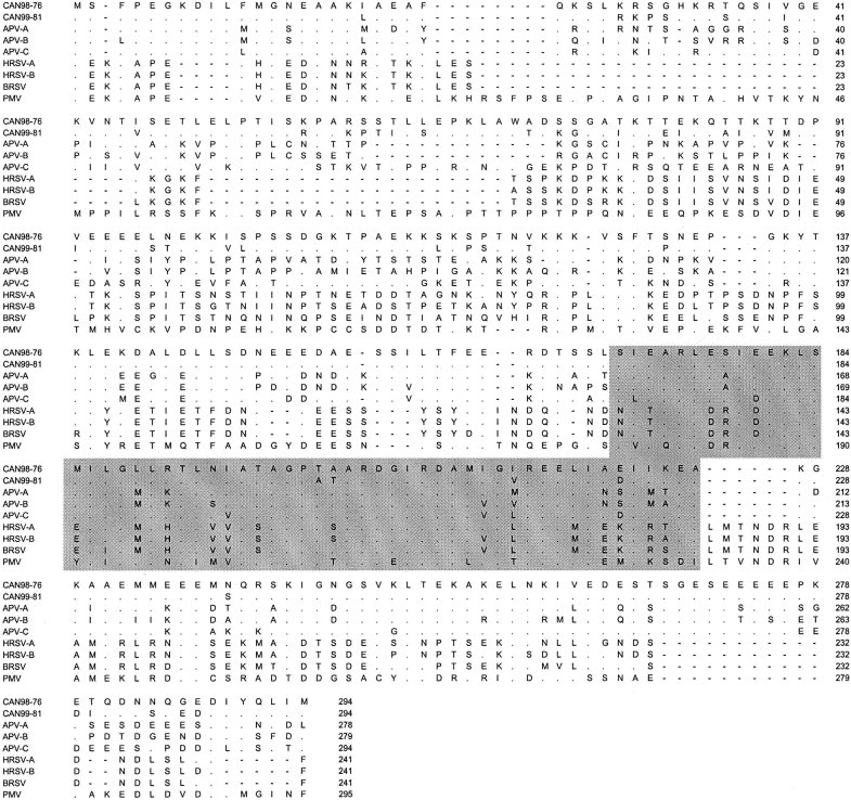
Alignment of the predicted amino acid sequences of the hMPV P protein of hMPV CAN99-81 (cluster 1) with those of CAN98-76 (cluster 2), APV-A, B and C, HRSV A and B, BRSV and PVM. Only residues that differ from isolate CAN98-76 are shown, identical amino acids are represented by periods, gaps are represented by dashes. A highly conserved domain in pneumovirus P (residues 171–226) is shaded.
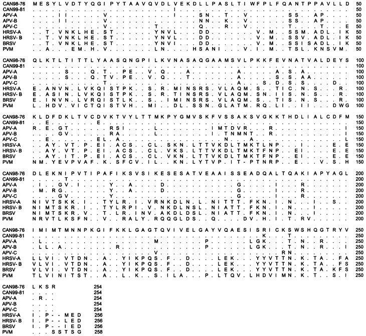
Alignment of the predicted amino acid sequences of the hMPV M protein of hMPV CAN99-81 (cluster 1) with those of CAN98-76 (cluster 2), APV-A, B and C, HRSV A and B, BRSV and PVM. Only residues that differ from isolate CAN98-76 are shown, identical amino acids are represented by periods, gaps are represented by dashes.
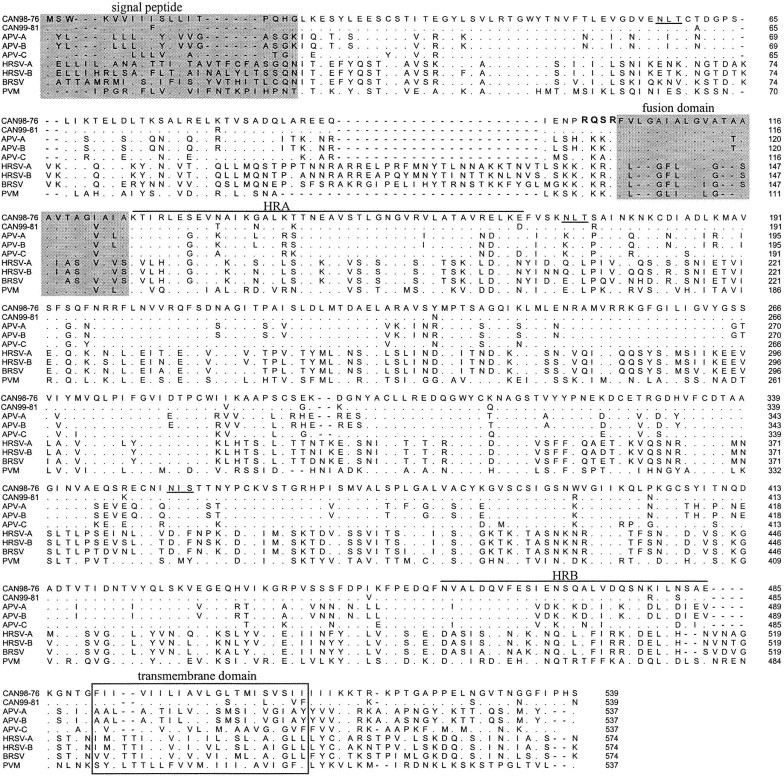
Alignment of the predicted amino acid sequences of the hMPV F protein of hMPV CAN99-81 (cluster 1) with those of CAN98-76 (cluster 2), APV-A, B and C, HRSV A and B, BRSV and PVM. Only residues that differ from isolate CAN98-76 are shown, identical amino acids are represented by periods, gaps are represented by dashes. The signal peptide and the fusion related domain sequences are shaded. The connecting peptide at the junction of the F1 and F2 subunit is bolded. The transmembrane domain is boxed. The two heptad repeats domains (HRA and HRB) are overlined The putative glycosylation sites present in the hMPV CAN98-76 sequence are underlined.
References
-
- Barr J., Chambers P., Pringle C.R., Easton A.J. Sequence of the major nucleocapsid protein gene of pneumonia virus of mice: sequence comparisons suggest structural homology between nucleocapsid proteins of pneumoviruses, paramyxoviruses, rhabdoviruses and filoviruses. J. Gen. Virol. 1991;72(Pt 3):677–685. - PubMed
-
- Bayon-Auboyer M.H., Arnauld C., Toquin D., Eterradossi N.’. Nucleotide sequences of the F, L and G protein genes of two non-A/non-B avian pneumoviruses (APV) reveal a novel APV subgroup. J. Gen. Virol. 2000;81:2723–2733. - PubMed
-
- Buckland R., Wild F.’. Leucine zipper motif extends. Nature. 1989;338:547. - PubMed
MeSH terms
Substances
LinkOut - more resources
Full Text Sources
Other Literature Sources
