. 2003 May 1;22(9):2234-44.
doi: 10.1093/emboj/cdg193.

Unified two-metal mechanism of RNA synthesis and degradation by RNA polymerase

Affiliations

Unified two-metal mechanism of RNA synthesis and degradation by RNA polymerase

Vasily Sosunov et al. EMBO J. .

Abstract

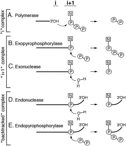
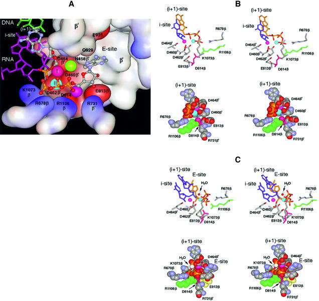
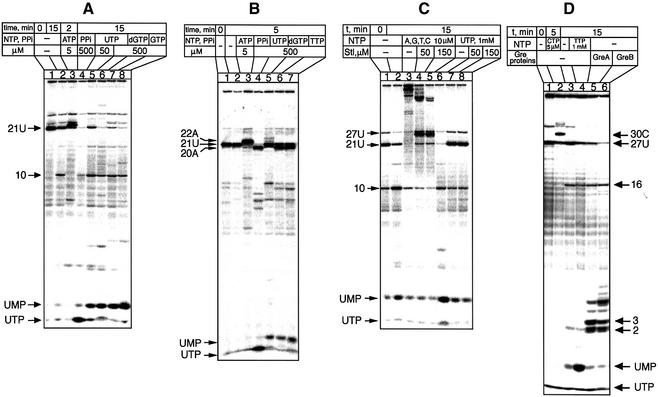
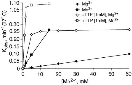
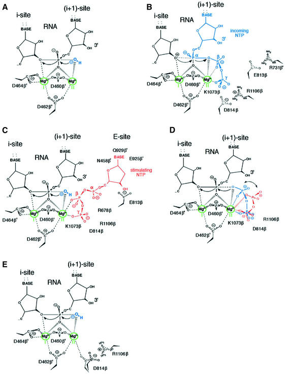
In DNA-dependent RNA polymerases, reactions of RNA synthesis and degradation are performed by the same active center (in contrast to DNA polymerases in which they are separate). We propose a unified catalytic mechanism for multisubunit RNA polymerases based on the analysis of its 3'-5' exonuclease reaction in the context of crystal structure. The active center involves a symmetrical pair of Mg(2+) ions that switch roles in synthesis and degradation. One ion is retained permanently and the other is recruited ad hoc for each act of catalysis. The weakly bound Mg(2+) is stabilized in the active center in different modes depending on the type of reaction: during synthesis by the beta,gamma-phosphates of the incoming substrate; and during hydrolysis by the phosphates of a non-base-paired nucleoside triphosphate. The latter mode defines a transient, non-specific nucleoside triphosphate-binding site adjacent to the active center, which may serve as a gateway for polymerization of substrates.


Figures

None
Fig. 1. Enzymatic activities of RNAP. Schematic representation of reactions performed by RNAP in various types of TEC. ‘i’ and ‘i + 1’ denote the two sites of the active center. Bold lines represent RNA. Bent arrows show the direction of nucleophilic attack.
None
Fig. 2. Molecular model of the RNAP active center. The model combines prior knowledge and present results, as explained in the text. (A) The active center in surface rendition performing the 3′–5′ exonuclease reaction. Basic and acidic amino acid residues are represented by the blue and red surfaces, respectively. In the top left of the panel, the 3′-terminal nucleoside (gray–blue metallic) in the i + 1 site is paired with the DNA base (green). The penultimate RNA nucleotide in the i site is depicted in purple. Of the four phosphates shown (yellow–red), one belongs to the 3′-terminal NMP in the i + 1 site and three belong to the non-complementary NTP in the E-site. Balls marked I and II represent catalytic Mg-I and Mg-II. Unmarked balls indicate the positions of Mg2+ ions from the published structures [green from Vassylyev et al. (2002); purple from Cramer et al. (2001)]. The water molecule participating in the exonuclease reaction is omitted. (B) Stereoscopic rendition (for crossed eyes) of the active center performing synthesis. Crucial amino acid residues are indicated. The purple and blue balls represent Mg-I and Mg-II, respectively. Top panel: the 3′-terminal nucleotide in the i site is shown in blue and the incoming NTP in the i + 1 site in yellow; its phosphates coordinate Mg-II. Bottom panel: space-filled model of the same reaction from a different angle. The 3′-terminal nucleotide is not shown; the incoming NTP in the i + 1 site is shown. (C) Stereo view of the active center performing the 3′–5 exonuclease reaction from two angles. The arrow points to the water molecule that serves as acceptor for nucleotidyl transfer. Top panel: the two 3′-terminal nucleotides (blue) are shown occupying the i and i + 1 sites, while the yellow NTP in the E-site coordinates Mg-II. Bottom panel: space-filled model from a different angle. Only the 3′-terminal nucleotide is shown in the i + 1 site.
None
Fig. 3. Characterization of the 3′–5′ exonuclease of RNAP. Autoradiograms show the effect of incubation of defined TECs carrying radiolabeled transcripts (lanes 1) under the indicated conditions. The RNA fragments are identified by their length (numbers) and 3′-terminal nucleotide (letters). The starting complexes in (A), (C) and (D) carry a 32P label only in the 3′-terminal nucleotide, while in (B) RNA was labeled both at the 3′-terminus and internally.
None
Fig. 4. Dependence of the 3′–5′ exonuclease reaction on Me2+ and TTP. The curves show the initial rate of UMP cleavage from 3′-terminally labeled TEC-21U as a function of Mg2+ or Mn2+ concentration in the presence or absence of 1 mM dTTP.
None
Fig. 5. Effect of mutations on the 3′–5′ exonuclease activity. (A) Autoradiograms show degradation of the terminally labeled transcript in TEC-21U by the wild-type and mutant RNAPs. (B) Dependence of the rate of UMP cleavage on Mg2+ concentration.
None
Fig. 6. Effect of pH on the 3′–5′ exonuclease and the pyrophosphorylase activities. (A and B) Autoradiograms show the release of terminal nucleotide from TEC-21U by the ED/AA mutant and wild-type RNAPs. (C) Effect of pH on the initial rate of terminal UMP release by the wild-type and mutant RNAPs at 10 mM MgCl2.
None
Fig. 7. Modes of the action of RNAP active center. The schemes present the results of molecular modeling and proposed reaction mechanisms when coordination of Mg-II is effected in different modes. (A) The 3′–5′ exonuclease in the absence of additional stabilization of Mg-II. (B) The polymerase with Mg-II stabilized by the incoming NTP (shown in blue). (C) The 3′–5′ exonuclease with Mg-II stabilized by non-complementary NTP (red). (D) The pyrophosphorylase with stabilizing pyrophosphate bound in two alternative modes (red or blue). (E) The 3′–5′ exonuclease with Mg-II stabilized by D814 residue shift resulting from disrupted interaction with R1106.

References

    1. Borukhov S. and Goldfarb,A. (1993) Recombinant Escherichia coli RNA polymerase: purification of individually overexpressed subunits and in vitro assembly. Protein Expr. Purif., 4, 503–511. - PubMed
    1. Borukhov S., Sagitov,V. and Goldfarb,A. (1993) Transcript cleavage factors from E.coli. Cell, 72, 459–466. - PubMed
    1. Brautigam C.A. and Steitz,T.A. (1998) Structural principles for the inhibition of the 3′–5′ exonuclease activity of Escherichia coli DNA polymerase I by phosphorothioates. J. Mol. Biol., 277, 363–377. - PubMed
    1. Cramer P., Bushnell,D.A. and Kornberg,R.D. (2001) Structural basis of transcription: RNA polymerase II at 2.8 Å resolution. Science, 292, 1863–1876. - PubMed
    1. Epshtein V., Mustaev,A., Markovtsov,V., Bereshchenko,O., Nikiforov,V. and Goldfarb,A. (2002) Swing-gate model of nucleotide entry into the RNA polymerase active center. Mol. Cell, 10, 623–634. - PubMed

Publication types