. 2003 May 1;22(9):2255-63.
doi: 10.1093/emboj/cdg206.

Functional interaction between PARP-1 and PARP-2 in chromosome stability and embryonic development in mouse

Affiliations

Functional interaction between PARP-1 and PARP-2 in chromosome stability and embryonic development in mouse

Josiane Ménissier de Murcia et al. EMBO J. .

Abstract

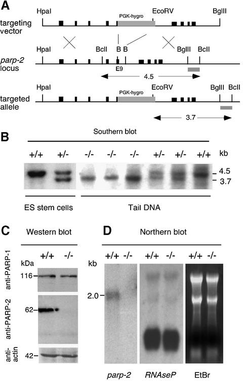
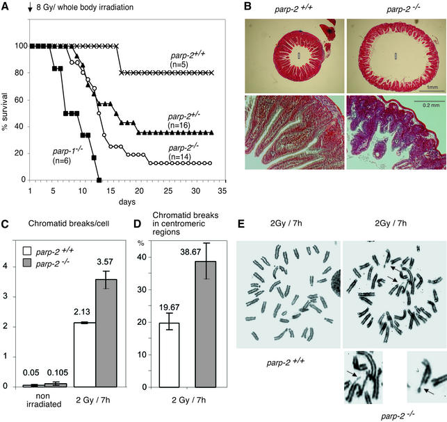
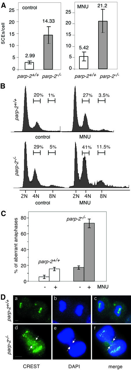
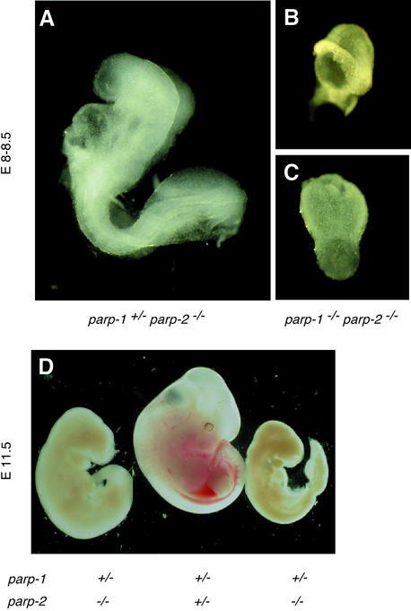
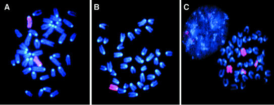
The DNA damage-dependent poly(ADP-ribose) polymerases, PARP-1 and PARP-2, homo- and heterodimerize and are both involved in the base excision repair (BER) pathway. Here, we report that mice carrying a targeted disruption of the PARP-2 gene are sensitive to ionizing radiation. Following alkylating agent treatment, parp-2(-/-)-derived mouse embryonic fibroblasts exhibit increased post-replicative genomic instability, G(2)/M accumulation and chromosome mis-segregation accompanying kinetochore defects. Moreover, parp-1(-/-)parp-2(-/-) double mutant mice are not viable and die at the onset of gastrulation, demonstrating that the expression of both PARP-1 and PARP-2 and/or DNA-dependent poly(ADP-ribosyl) ation is essential during early embryogenesis. Interestingly, specific female embryonic lethality is observed in parp-1(+/-)parp-2(-/-) mutants at E9.5. Meta phase analyses of E8.5 embryonic fibroblasts highlight a specific instability of the X chromosome in those females, but not in males. Together, these results support the notion that PARP-1 and PARP-2 possess both overlapping and non-redundant functions in the maintenance of genomic stability.


Figures

None
Fig. 1. Targeted disruption of parp-2. (A) Map of the parp-2 murine genomic locus and targeting vector. Exons are indicated by black boxes and the position of the external probe is represented by a gray box. The PGK-hygromycin cassette is inserted in the opposite transcriptional orientation and replaces a BamHI (B) fragment of exon 9 and intron 10. (B) Representative genomic blot probed with the external probe. A wild-type 4.5 kb and a recombined 3.7 kb fragment are shown. Mice were identified as wild-type (+/+), heterozygous (+/–) or homozygous mutant (–/–). (C) PARP-2 is not expressed in cells isolated from the testis of parp-2–/– mice. (D) Northern blot analysis of total RNA from testis using a 820 bp EcoRI fragment of parp-2 cDNA as a probe. Note that the expression of the RNase P RNA is not influenced by the parp-2 disruption.
None
Fig. 2. parp-2–/– mice are sensitive to ionizing radiation. (A) Kaplan–Meier survival curve after 8 Gy of irradiation. Wilcoxon test: P (parp-2+/+ versus parp-2–/–) < 10–5; P (parp-2–/– versus parp-2+/–) < 0.002. (B) Stained transverse sections of the duodenum from a parp-2+/+ and a parp-2–/– mouse 6 days after a dose of 8 Gy was given. Lumen (l), villi (v). (C) Mean number of chromatid breaks in bone marrow cells taken from mock-irradiated mice or 7 h after 2 Gy whole-body irradiation. (D) Occurrence of chromatid breaks in the centromeric region 7 h after 2 Gy irradiation. (E) Metaphase spreads of bone marrow cells from irradiated parp-2+/+ and parp-2–/– mice. Chromatid break in the centromeric region (top arrow) and tetraradian in metaphase (bottom arrow) from an irradiated parp-2–/– mouse. Magnifications are details pointed to by arrows.
None
Fig. 3. (A) Sister chromatid exchanges in bone marrow cells from parp-2+/+ and parp-2–/– mice 9 h after i.p injection with (50 mg/kg) MNU. (B) Cell cycle profile analyzed after propidium iodide staining of parp-2+/+ and parp-2–/– MEFs at passage 2, 24 h following 2 mM MNU treatment. The percentage of cells containing (2N) (4N) and (8N) is indicated. (C) Quantitative analysis of chromosome mis-segregation in parp-2+/+ and parp-2–/– MEFs at passage 2, 24 h following 2 mM MNU treatment. The fraction of mitotic cells that exhibited a defect in segregation, such as lagging chromosomes (see D, panels d–f), is shown. (D) Anaphases in parp-2+/+ and parp-2–/– MEFs following 2 mM MNU treatment. Kinetochores (green) are immunostained with the CREST serum (a and d). Lagging chromosomes forming an anaphase bridge indicated by arrows in parp-2–/– cells. Bars indicate 10 µm.
None
Fig. 4. (A) Fluorescent TUNEL stain of thymus 2 days after 6 Gy irradiation. Apoptotic cells are labeled in green, and cell nuclei are stained with DAPI. parp +/+ are sparsely stained whereas parp-1–/– and parp-2–/– show a strong green staining of apoptotic cells. (B) PARP-1 and PARP-2 cleavage kinetics after 100 µM VP16 treatment in HL-60 cells. Z-VAD-fmk inhibitor was used at 0.5 µg/µl and was added 1 h before VP16 treatment. For western blot, 105 cells were harvested at the indicated time, mixed with loading buffer and sonicated. Polyclonal antibodies anti-PARP-1 and anti-PARP-2 were used. (CIn vitro cleavage of purified mPARP-2 (2.5 µg) by active caspase-3 (8 U, Chemicon International) shows the same cleavage pattern in HL-60 cells after VP-16 treatment. The N-terminus of the 55 kDa band was micro sequenced and was found to be 58DNRD61, a consensus cleavage site for caspases 3/7.
None
Fig. 5. External views of E8.0–8.5 (A–C) and E11.5 (D) embryos. (A) A normal parp-1+/–parp-2–/– embryo. (B and C) Retarded parp-1–/– parp-2–/– embryos. (D) Example of two parp-1+/–parp-2–/– female embryos that are phenotypically arrested at E9.5, and one normal parp-1+/–parp-2+/– female embryo at E11.5. The same magnifications were used in (A), (B) and (C).
None
Fig. 6. parp-1+/–parp-2–/– females display X-chromosome instability. X chromosomes are stained in pink (rhodamine), and chromosomes are counterstained in blue (DAPI). Examples of metaphases with various numbers of X chromosomes in parp-1+/–parp-2–/– females: (A) 2X; (B) 1X; (C) 2X + derX.

References

    1. Allen J.W., Shuler,C.F., Mendes,R.W. and Latt,S.A. (1977) A simplified technique for in vivo analysis of sister-chromatid exchanges using 5-bromodeoxyuridine tablets. Cytogenet. Cell Genet., 18, 231–237. - PubMed
    1. Amé J.C. et al. (1999) PARP-2, a novel mammalian DNA damage-dependent poly(ADP-ribose) polymerase. J. Biol. Chem., 274, 17860–17868. - PubMed
    1. Amé J.C., Schreiber,V., Fraulob,V., Dollé,P., de Murcia,G. and Niedergang,C.P. (2001) A bidirectional promoter connects the poly(ADP-ribose) polymerase 2 (PARP-2) gene to the gene for RNase P RNA. Structure and expression of the mouse PARP-2 gene. J. Biol. Chem., 276, 11092–11099. - PubMed
    1. Avner P. and Heard,E. (2001) X-chromosome inactivation: counting, choice and initiation. Nat. Rev. Genet., 2, 59–67. - PubMed
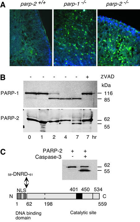
    1. Benchoua A., Couriaud,C., Guegan,C., Tartier,L., Couvert,P., Friocourt,G., Chelly,J., Menissier-de Murcia,J. and Onteniente,B. (2002) Active caspase-8 translocates into the nucleus of apoptotic cells to inactivate poly(ADP-ribose) polymerase-2. J. Biol. Chem., 277, 34217–34222. - PubMed

Publication types