

Comment
doi: 10.1172/JCI18539.
Double target for tumor mass destruction
Affiliations
- PMID: 12727916
- PMCID: PMC154456
- DOI: 10.1172/JCI18539
Item in Clipboard
Comment
Double target for tumor mass destruction
J Clin Invest.
2003 May.
No abstract available
Figures
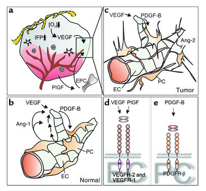
Growth factor receptor signals involved in tumor angiogenesis and vascular development. A solid tumor is dependent on new blood vessels for growth. (a) Angiogenesis is induced by VEGF produced mainly by hypoxic tumor cells and inflammatory cells (such as macrophages, which also secrete MMPs involved in the angiogenic switch) (19). Placental growth factor (PlGF) produced in the tumor may recruit bone marrow–derived endothelial precursor cells (EPCs), which become incorporated into the angiogenic vessels (20). The tip cells of a growing angiogenic sprout, and also the tumor cells, secrete PDGF-B, which binds to the EC surface and recruits PCs to migrate along the angiogenic sprouts (8, 9). In normal tissues, blood vascular ECs and PCs are in tight contact, and angiopoietin-1 (Ang-1) increases the stability of the vessels (b). In tumor vessels, Ang-2, an antagonist of Ang-1, is produced by the ECs, and the PCs are often only loosely attached to EC tubes; they also have long projections reaching out from the vessels to the surrounding matrix (13, 22) (c). The tumor blood vessels are leaky and the peritumoral lymphatic vessels (shown in yellow in panel a) cannot clear the extravasated material (10), which results in increased interstitial fluid pressure (IFP) within the tumor. The findings of Bergers et al. now suggest that concomitant inhibition of ECs and PCs through inhibition of their respective transmembrane tyrosine kinase receptors VEGFR-2 and VEGFR-1 (d) and PDGFR-β (e) results in more efficient inhibition of angiogenesis (4). The roles of additional ligands, VEGF-B (23) and PDGF-D (24, 25), which bind to VEGFR-1 and PDGFR-β, respectively, are not yet clear in these processes.
Comment on
-
Benefits of targeting both pericytes and endothelial cells in the tumor vasculature with kinase inhibitors.J Clin Invest. 2003 May;111(9):1287-95. doi: 10.1172/JCI17929. J Clin Invest. 2003. PMID: 12727920 Free PMC article.
References
-
- Folkman J. Role of angiogenesis in tumor growth and metastasis. Semin. Oncol. 2002;29:15–18. - PubMed
-
- Pugh CW, Ratcliffe PJ. The von Hippel-Lindau tumor suppressor, hypoxia-inducible factor-1 (HIF-1) degradation, and cancer pathogenesis. Semin. Cancer Biol. 2003;13:83–89. - PubMed
-
- Alitalo K, Carmeliet P. Molecular mechanisms of lymphangiogenesis in health and disease. Cancer Cell. 2002;1:219–227. - PubMed
-
- Benjamin LE, Hemo I, Keshet E. A plasticity window for blood vessel remodeling is defined by pericyte coverage of the preformed endothelial network and is regulated by PDGF-B and VEGF. Development. 1998;125:1591–1598. - PubMed
Publication types
MeSH terms
Substances
LinkOut - more resources
Full Text Sources
Other Literature Sources
