. 2003 May 15;257(2):263-77.
doi: 10.1016/s0012-1606(03)00068-x.

dHAND-Cre transgenic mice reveal specific potential functions of dHAND during craniofacial development

Affiliations

dHAND-Cre transgenic mice reveal specific potential functions of dHAND during craniofacial development

Louis-Bruno Ruest et al. Dev Biol. .

Abstract

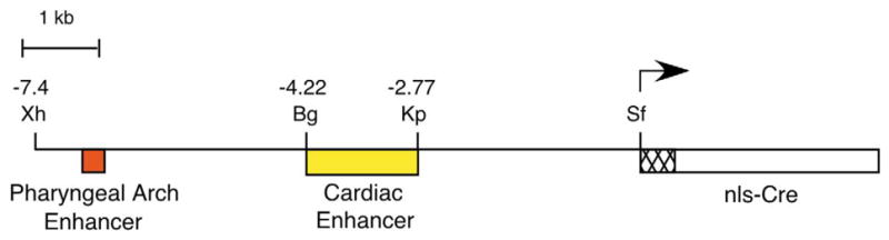
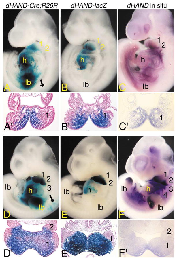
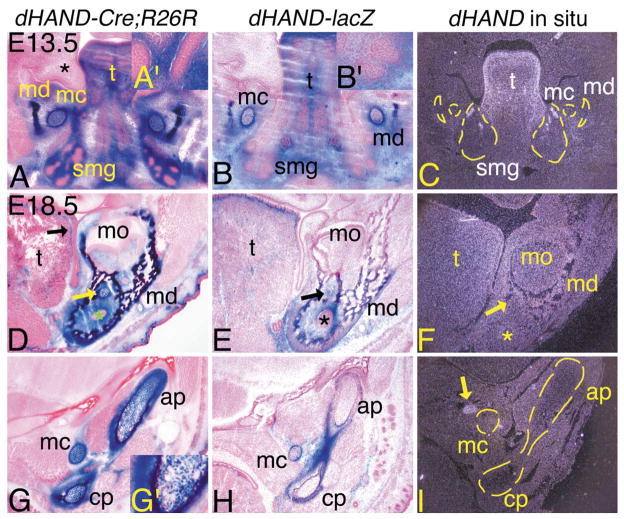
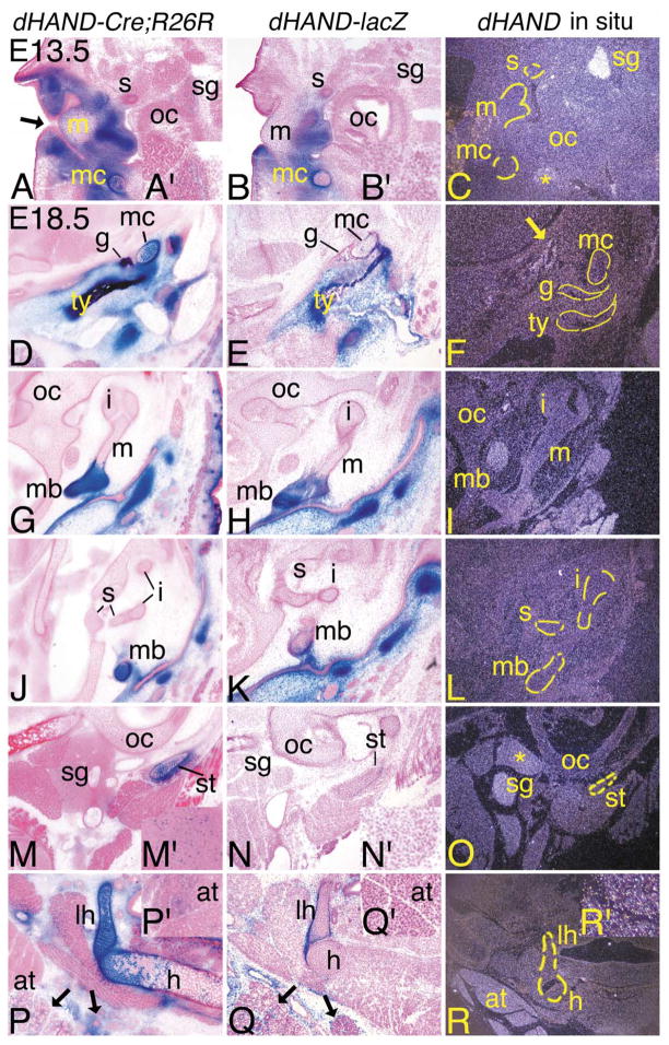
Most of the bone, cartilage, and connective tissue of the craniofacial region arise from cephalic neural crest cells. Presumably, patterning differences in crest cells are a result of regional action of transcription factors within the developing pharyngeal arches. The basic helix-loop-helix transcription factor dHAND/HAND2 is expressed throughout much of the neural crest-derived mesenchyme of the pharyngeal arches, suggesting that it plays a crucial role in craniofacial development. However, targeted inactivation of the dHAND gene results in embryonic lethality by E10.5 due to vascular defects, preventing further analysis of the role of dHAND in cephalic neural crest cell development. In order to examine putative roles of dHAND during later stages of embryogenesis, we have used a transgenic lineage marker approach, in which a portion of the dHAND upstream region containing an enhancer that directs dHAND expression to the pharyngeal arches is used to drive Cre recombinase expression. By crossing these dHAND-Cre transgenic mice with R26R mice, we can follow the fate of cells that expressed dHAND at any time during development by examining beta-galactosidase activity. We show that dHAND is first expressed in postmigratory cephalic neural crest cells within the pharyngeal arches. In older embryos, beta-galactosidase-labeled cells are observed in most of the neural crest-derived lower jaw skeleton and surrounding connective tissues. However, labeled cells only contribute to substructures within the middle ear, indicating that our transgene is not globally expressed in cephalic neural crest cells within the pharyngeal arches. Moreover, dHAND-Cre mice will provide a valuable tool for tissue-specific inactivation of gene expression in multiple tissue types of neural crest origin.


Figures

Fig. 1
Fig. 1
Diagram of the dHAND-Cre transgene. A 7.4-kb fragment of the dHAND upstream region, containing enhancers required for pharyngeal arch and cardiac expression, was fused to a Cre cDNA containing nuclear localization and SV40 polyadenylation sequences. A synthetic intron resides in the 5′ end of the Cre cDNA to aid transgene expression. Bg, BglII; Kp, KpnI; Sf, SfiI; Xh, XhoI.
Fig. 2
Fig. 2
Early dHAND expression domains in dHAND-Cre; R26R embryos. (A–F) Lateral (A–F) and transverse (A′–F′) views of dHAND-Cre;R26R (A, A′; D, D′) and dHAND-lacZ (B, B′; E, E′) embryos stained in whole mount for β-gal activity and wild type embryos (C, C′; F, F′) after whole-mount in situ hybridization analysis of dHAND expression. Transverse sections of β-gal-stained embryos were counterstained with nuclear fast red and photographed by using brightfield optics. Transverse sections of whole-mount in situ embryos were photographed by using Nomarski DIC optics. (A) In E9.5 dHAND-Cre; R26R embryos, labeled cells are observed in the first mandibular (1) and second (2) arches, common heart ventricle (h), and forelimb bud (lb). Few scattered cells are observed in the early dorsal root ganglion (black arrow). (A′) A transverse section through the first arch of the embryo shown in (A) illustrates that stained cells are confined to the arch mesenchyme in a dHAND-specific pattern. (B) In E9.5 dHAND-lacZ embryos, staining is observed in the mandibular arch and common heart ventricle. (B′) A transverse section through the first arch of the embryo shown in (B) shows that labeled cells are confined to the arch mesenchyme. (C) In E9.5 wild type embryos, dHAND expression is observed in the first mandibular and second arches and in the future right ventricle of the heart (obscured by the left ventricle). (C′) As observed in (A′) and (B′), dHAND expression is confined to the arch mesenchyme. (D) By E10.5, labeled cells in dHAND-Cre;R26R embryos are observed in arches 1–3, the ventricle, and the limb bud. Scattered cells are also observed in the atrium. While not visible in this view, stained cells are also observed in arch 4. Labeled cells are also observed in the cervical dorsal root ganglion (black arrows). (D′) In a transverse section through the first and second arches, staining is observed in the arch mesenchyme, with the intensity of staining highest in the periphery of the arch. (E) In E10.5 dHAND-lacZ embryos, labeled cells are observed in arches one and two and in the ventricles of the heart. Scattered cells are also present in the atria. (E′) As observed in dHAND-Cre;R26R embryos, staining within the mesenchyme of the first arch is strongest at the periphery. While not shown due to a slight difference in the angle of section, the second arch staining in dHAND-lacZ embryos is comparable with that of dHAND-Cre;R26R embryos. (F) dHAND expression in E10.5 wild type embryos is observed in arches 1– 4, distal limb buds, and right ventricle of the heart. (F′) A section through the first and second arch again illustrates staining throughout the medial half of the arch mesenchyme, with the strongest staining observed along the periphery of the arch.
Fig. 3
Fig. 3
Contribution of dHAND lineage cells to arch-derived structures of the jaw of dHAND-Cre;R26R embryos. Transverse (A–C) and frontal (D–I) sections through the heads of dHAND-Cre;R26R (A, D, G), dHAND-lacZ (B, E, H), and wild type (C, F, I) embryos. Frozen sections from dHAND-Cre;R26R and dHAND-lacZ embryos were stained for β-gal activity and counterstained with nuclear fast red (E13.5) or eosin (E18.5). Paraffin sections from wild type embryos were probed with a 35S-labeled riboprobe against dHAND. After developing, sections were counterstained with Harris hematoxylin and photographed under darkfield conditions. To aid in the visualization of specific structures, yellow lines have been added to some of the darkfield panels. (A–C) Transverse sections through the lower jaw of E13.5 embryos. (A) Labeled cells in dHAND-Cre;R26R embryos are observed in structures derived from the first pharyngeal arch, including mandibular bone (md), Meckel’s cartilage (mc), tongue (t), and surrounding mesenchyme/connective tissue. Abundant labeled cells are also observed in the mesenchyme of the developing submandibular glands (smg), but not in the glandular epithelium (see inset, A′). The mesenchyme of the upper jaw (future maxilla; *) and oral cavity epithelium do not contain labeled cells. (B) Labeled cells in dHAND-lacZ transgenic embryos are observed in a similar pattern to those in dHAND-Cre;R26R embryos, though labeled cells in Meckel’s cartilage are mixed with unlabeled cells. (C) Endogenous dHAND expression is observed in the lamina propria of the tongue and in the mesenchyme surrounding the proximal ducts of the submandibular gland. Very little expression is observed in the mandible or Meckel’s cartilage. (D–I) Frontal sections through the mandible of E18.5 embryos. (D) β-gal-stained cells in dHAND-Cre;R26R embryos are present in the connective tissue of the tongue (t), with highest staining in the lamina propria (black arrow). Almost all cells in the mandible, Meckel’s cartilage (yellow arrow), and surrounding mesenchyme are also labeled. (E) Labeled cells in dHAND-lacZ embryos are again observed in a similar pattern to those in dHAND-Cre;R26R embryos. However, a mixture of labeled and unlabeled cells composes the mandibular bone and Meckel’s cartilage. (F) Faint dHAND expression is observed in the mandibular bone. Very little expression is observed in Meckel’s cartilage. The asterisks in (D–F) mark the enamel organ of the left incisor. (G) In a more proximal section to that shown in (D–F), labeled cells in dHAND-Cre;R26R embryos are observed in Meckel’s cartilage and in the articular (ap) and coronoid (cp) processes of the mandible and surrounding perichondrium. However, fewer labeled cells are observed near the junction with the ossified mandible, an area undergoing endochondral ossification and composed of hypertrophic chondrocytes (see inset, G′). (H) Labeled cells are mixed with unlabeled cells in Meckel’s cartilage, but are not observed in the anterior and coronoid processes of the mandible. The perichondrium surrounding the process is composed almost solely of labeled cells. (I) Endogenous dHAND expression is not observed in the mandibular processes or in Meckel’s cartilage, though expression is observed in the sympathetic ganglion (yellow arrow). Due to the angle of section, this structure is not observed in the dHAND-Cre-R26R or dHAND-lacZ embryo sections, though scattered labeled cells are present in the sympathetic ganglion of dHAND-Cre-R26R embryos (see Fig. 4). mo, molar.
Fig. 4
Fig. 4
Distribution of dHAND progeny cells in the middle ear and throat during development of dHAND-Cre; R26R embryos. Transverse (A–C; E13.5) and frontal (D–R; E18.5) sections through the middle ear and throat of dHAND-Cre;R26R (A, D, G, J, M, P), dHAND-lacZ (B, E, H, K, N, Q), and wild type (C, F, I, L, O, R) embryos. Treatment of sections was performed as described in Fig. 3. (A–C) Sections through the middle ear of E13.5 embryos. (A) In dHAND-Cre;R26R embryos, stained cells are observed in the mesenchyme surrounding the epithelium of the external auditory meatus (black arrow), in Meckel’s cartilage (mc), and in the future manubrium of the malleus (m). Scattered labeled cells are also observed in the sympathetic ganglion (sg; see inset, A′). Labeled cells are not observed in the stapes (s), though are present in more proximal sections (see text). (B) Labeled cells in dHAND-lacZ embryos are also observed in the mesenchyme of the middle ear region, the manubrium of the malleus, and Meckel’s cartilage, though they are mixed with unlabeled cells in Meckel’s cartilage. No staining is observed in the stapes or sympathetic ganglion (see inset, B′). (C) Very little endogenous dHAND expression is observed in the middle ear. The exceptions are the sympathetic ganglion and the mandibular branch of the trigeminal ganglion (*). (D–R) Frontal sections through the heads of E18.5 embryos. (D) The tympanic (ty) and gonial (g) bones and Meckel’s cartilage are composed almost solely of labeled cells in dHAND-Cre;R26R embryos. The surrounding mesenchyme in this region of the head is also composed of labeled cells. (E) Labeled cells in dHAND-lacZ embryos are observed in the tympanic ring and gonial bone, though a small portion of unlabeled cells are also present. More significant mixing of labeled and unlabeled cells is observed in Meckel’s cartilage. (F) Little endogenous expression is observed in the tympanic and gonial bones and Meckel’s cartilage, though faint expression is found along some of the head vasculature (arrow). (G, H) In sections through the malleus, labeled cells in dHAND-Cre;R26R and dHAND-lacZ embryos are confined to the manubrium (mb) of the malleus and surrounding mesenchyme. (I) dHAND expression is not observed in any part of the malleus or incus (i) in wild type embryos. (J, K) β-gal-stained cells in dHAND-Cre;R26R and dHAND-lacZ embryos are not present in the incus or stapes (s). Labeled cells in the manubrium of dHAND-lacZ embryos are mixed with unstained cells. (L) dHAND expression is not observed in the incus, stapes, or manubrium of the malleus. (M) In sections through the distal portion of the styloid cartilage (st), labeled cells in dHAND-Cre;R26R embryos compose most of the styloid and are scattered in the sympathetic ganglion (see inset, M′). (N) In dHAND-lacZ embryos, labeled cells are present in the perichondrium of the styloid cartilage, but are mixed in the cartilage itself and are absent in the sympathetic ganglion (see inset, N′). (O) dHAND expression is observed in the sympathetic ganglion in the incus/stapes region, with weaker expression in the glossopharyngeal ganglion. (P) In the throat, β-gal-stained cells in dHAND-Cre;R26R embryos are observed in the lesser horn (lh) and body (h) of the hyoid. However, labeled cells are mixed with unlabeled cells in the body, possibly reflecting changes in staining in cartilage undergoing endochondral ossification, as few hypertrophic chondrocytes are stained. Labeled cells are also observed in the mesenchyme of the submandibular and sublingual glands (black arrows). Out of the photographic frame, adipose tissue also contains scattered labeled cells (inset, P′). (Q) As with other cartilages, labeled cells in dHAND-lacZ embryos compose the perichondrium of the lessor horns of hyoid but are mixed with a majority of unlabeled cells in the cartilage itself. Very few labeled cells are observed in the body of the hyoid. Labeled cells are observed in the mesenchyme of the submandibular and sublingual glands (arrows), but are absent in adipose tissue observed out of the photographic frame (inset, Q′). (R) dHAND expression is not observed in or around the hyoid cartilages or bones; however, adipose tissue at the base of the neck shows strong dHAND expression. Out of the plane of section, expression is also observed in the submandibular gland (inset, R′). oc, otic capsule.
Fig. 5
Fig. 5
Contribution of dHAND progeny cells during odontogenesis in dHAND-Cre;R26R embryos. Transverse (A–L) and frontal (M–R) sections through the heads of dHAND-Cre;R26R (A, D, G, J, M, P), dHAND-lacZ (B, E, H, K, N, Q), and wild type (C, F, I, L, O, R) embryos. Treatment of sections was performed as described in Fig. 3. (A–C) Transverse sections through the incisors of E13.5 embryos. (A, B) Labeled cells in dHAND-Cre;R26R and dHAND-lacZ embryos are apparent throughout the developing lower jaw, including in Meckel’s cartilage (mc) and mesenchyme destined to become dentin and pulp of the lower incisors. Labeled cells are not observed in the oral epithelium (oep) or developing enamel organ (eo). (C) dHAND expression in wild type embryos is observed in the tongue (t) and mesenchyme surrounding the incisor enamel organs. Less expression is observed in Meckel’s cartilage. (D–F) Transverse sections through the molar of E13.5 embryos, showing developing dentition of the molar at the late bud stage. (D) Labeled cells in dHAND-Cre;R26R embryos are observed in the mesenchyme surrounding the bud of the developing mandibular molar (mdm; arrows) and mandibular bone (md). Oral epithelium and developing enamel organs (*) of both maxillary (mxm) and mandibular molars are unlabeled. (E) Labeled cells in dHAND-lacZ embryos are not observed surrounding the mandibular molar buds (arrow). (F) Endogenous dHAND expression is also not observed surrounding the mandibular molar buds (arrow). (G–I) Transverse sections through the incisors of E15.5 embryos. The incisors have progressed to a cap stage, approaching the “bell” configuration. (G) The nonlabeled enamel organ in dHAND-Cre;R26R embryos partially encases the labeled dental papilla (dp) and appears irregular in shape due to the transverse angle of the section. The symphysis of Meckel’s cartilage is almost completely composed of labeled cells. (H) Labeled cells in dHAND-lacZ embryos also compose most of the dental pulp of the incisor, in contrast to the mixing of labeled and unlabeled cells in the symphysis of Meckel’s cartilage. (I) dHAND expression is observed in the dental pulp and surrounding mesenchyme. Expression is much weaker in Meckel’s cartilage symphysis. (J–L) Transverse sections through the molar of E15.5 embryos. (J) In the mandibular molar, also in the cap stage, labeled cells in dHAND-Cre; R26R embryos contribute to the dental papilla (arrow), though this area also contains unlabeled cells. (K) The dental papilla of mandibular molars from dHAND-lacZ embryos is composed solely of unlabeled cells (arrow). (L) dHAND expression is not observed in the papilla of the mandibular molars (white arrow), in contrast to the strong expression in the lamina propria of the tongue. (M–O) Frontal sections through the incisors of E18.5 embryos. (M) Labeled cells in dHAND-Cre;R26R embryos appear in the dental papilla of the mandibular incisors, mandibular bone (md), Meckel’s cartilage, and surrounding mesenchyme. Oral epithelium remains unlabeled, as does the developed enamel matrix (em). (N) A similar pattern of staining is observed in the incisors of dHAND-lacZ embryos, though few labeled cells are observed in Meckel’s cartilage. (O) dHAND expression is observed in the incisor dental pulp, surrounding bone, and mesenchyme. Very little expression is observed in Meckel’s cartilage. (P–R) Frontal sections through the molars of E18.5 embryos. (P) Labeled cells in dHAND-Cre;R26R embryos appear in the dental papilla, although extensive mixing with unlabeled cells has occurred. Labeled cells are also observed in the mandibular bone, Meckel’s cartilage, and surrounding connective tissue. The residual dental lamina (dl) can be seen connecting the unlabeled oral epithelium and enamel organ. Labeled cells are not observed in mandibular nerves (arrow). (Q) Labeled cells in dHAND-lacZ embryos are confined to the mandibular bone and Meckel’s cartilage. Labeled cells are not observed in the dental papilla of the molar. (R) A low level of dHAND expression is observed in the mandibular bone surrounding the molar of wild type embryos, but expression is not observed in the molar dental pulp.

Similar articles

Cited by

References

    1. Bachmair A, Finley D, Varshavsky A. In vivo half-life of a protein is a function of its amino-terminal residue. Science. 1986;234:179–186. - PubMed
    1. Bronner-Fraser M. Origins and developmental potential of the neural crest. Exp Cell Res. 1995;218:405–417. - PubMed
    1. Carvajal JJ, Cox D, Summerbell D, Rigby PWJ. A BAC transgenic analysis of the Mrf4/Myf5 locus reveals interdigitated elements that control activation and maintenance of gene expression during muscle development. Development. 2001;128:1857–1868. - PubMed
    1. Chai Y, Jiang X, Ito Y, Bringas P, Han J, Rositch DH, Soriano P, McMahon AP, Sucov HM. Fate of the mammalian cranial neural crest during tooth and mandibular morphogenesis. Development. 2000;127:1671–1679. - PubMed
    1. Charité J, McFadden DG, Merlo GR, Levi G, Clouthier DE, Yanagisawa M, Richardson JA, Olson EN. Role of Dlx6 in regulation of an endothelin-1-dependent, dHAND branchial arch enhancer. Genes Dev. 2001;15:3039–3049. - PMC - PubMed

Publication types

MeSH terms

LinkOut - more resources