. 2003 May 1;17(9):1141-52.
doi: 10.1101/gad.1070003.

A novel ring-like complex of Xenopus proteins essential for the initiation of DNA replication

Affiliations

A novel ring-like complex of Xenopus proteins essential for the initiation of DNA replication

Yumiko Kubota et al. Genes Dev. .

Abstract

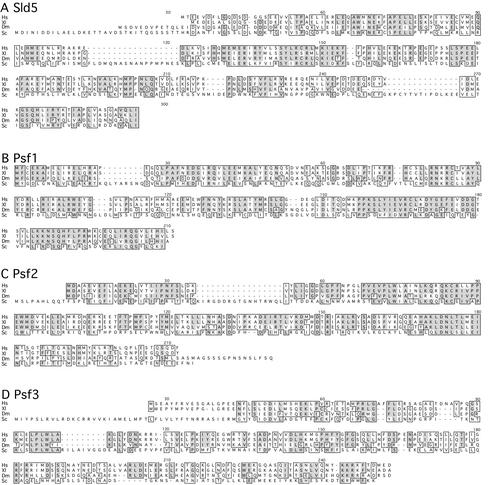
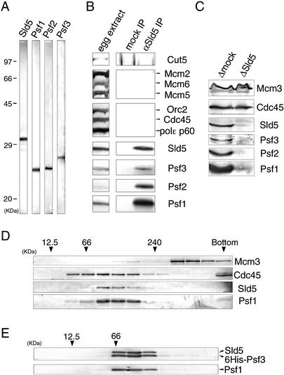
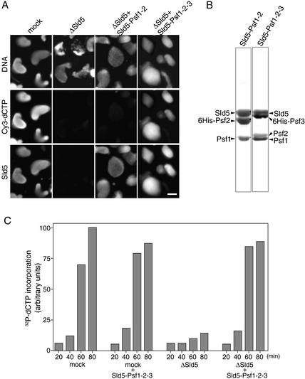
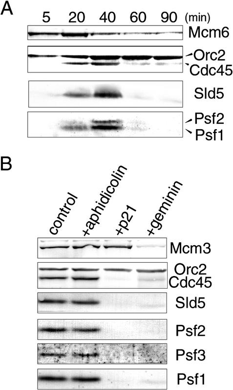
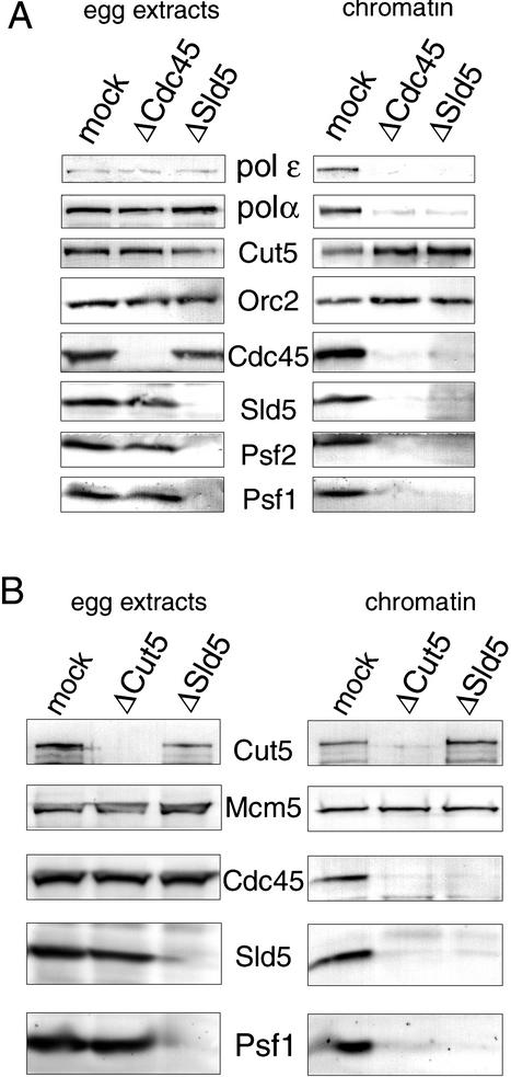
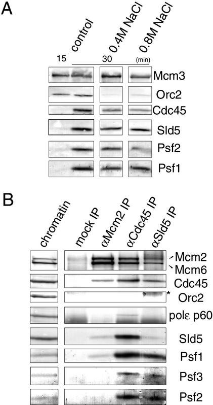
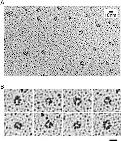
We have identified Xenopus homologs of the budding yeast Sld5 and its three interacting proteins. These form a novel complex essential for the initiation of DNA replication in Xenopus egg extracts. The complex binds to chromatin in a manner dependent on replication licensing and S-phase CDK. The chromatin binding of the complex and that of Cdc45 are mutually dependent and both bindings require Xenopus Cut5, the yeast homolog of which interacts with Sld5. On replicating chromatin the complex interacts with Cdc45 and MCM, putative components of replication machinery. Electron microscopy further reveals that the complex has a ring-like structure. These results suggest that the complex plays an essential role in the elongation stage of DNA replication as well as the initiation stage.


Figures

Figure 1
Figure 1
Alignment of predicted amino acids sequences of Sld5 (A), Psf1 (B), Psf2 (C), and Psf3 (D) from human (Hs), Xenopus (Xl), Drosophila (Dm), and Saccharomyces cerevisiae (Sc). Identical amino acids are shaded, and highly conserved regions are boxed.
Figure 2
Figure 2
Xenopus Sld5, Psf1, Psf2, and Psf3 form a complex in Xenopus egg extracts. (A) Specificity of antibodies. S-phase egg extract was resolved with SDS-PAGE and immunoblotted with the antibodies indicated in the figure. (B,C) Immunoprecipitation and immunodepletion of Sld5 from egg extracts. S-phase egg extracts were treated with preimmune (mock) or anti-Sld5 antibodies conjugated to protein A beads. The immunoprecipitates (B) and the depleted extracts (C) were resolved by SDS-PAGE, blotted, and probed with antibodies against the various proteins indicated in the figure. (D,E) Separation of proteins by glycerol gradient centrifugation. Soluble fractions of egg extracts (D) or purified complex of recombinant Sld5–Psf1–Psf2–Psf3 (E) were analyzed on glycerol gradients. Fractions were resolved by SDS-PAGE, blotted, and probed with antibodies against the proteins indicated in the figure. Marker proteins were centrifuged under identical conditions; peak positions of the marker proteins are indicated by arrowheads with their molecular weights.
Figure 3
Figure 3
Requirement of GINS for DNA replication in Xenopus egg extracts. (A) Immunofluorescent detection of nuclear Sld5. Xenopus sperm chromatin was incubated in the presence of Cy3-dCTP in mock- or Sld5-depleted extracts supplemented with or without recombinant Sld5–Psf1–Psf2 or Sld5–Psf1–Psf2–Psf3 (GINS). After incubation at 23°C for 45 min, samples were fixed and centrifuged through 30% sucrose onto coverslips. Nuclear localization of Sld5 was visualized with rabbit anti-Xenopus-Sld5 antibody followed by Alexa488-labeled anti-rabbit IgG. DNA replication was monitored as the incorporation of Cy3-dCTP into DNA, and DNA was visualized with Hoechst 33258 dye. Bar, 10 μm. (B) Protein compositions of recombinant Xenopus Sld5–Psf1–Psf2 and Sld5–Psf1–Psf2–Psf3 complexes. Xenopus Sld5, Psf1, and Psf2 were coexpressed with and without Psf3 (either Psf2 or Psf3 was tagged with His6) in Sf9 cells using a baculovirus expression system. The protein complexes, purified with Ni-NTA resin, were resolved by SDS-PAGE and stained with Ponceau S. (C) Replication activities of Sld5-depleted extracts. Xenopus sperm chromatin was incubated in mock- or Sld5-depleted extracts in the presence of 32P-labeled dCTP, supplemented with or without recombinant GINS. At the indicated times, DNA was isolated and subjected to alkali agarose gel electrophoresis followed by autoradiography. The amount of 32P incorporated into DNA was quantified and expressed in arbitrary units.
Figure 4
Figure 4
Requirement of replication licensing and S-CDK activity for the chromatin binding of Xenopus GINS. (A) The time course of chromatin binding of replication-related proteins. Sperm chromatin was incubated in egg extracts, and samples were collected at the indicated times. Chromatin fractions isolated by centrifugation were resolved by SDS-PAGE and immunoblotted with antibodies as indicated in the figure. (B) Effect of inhibitors of DNA replication on the chromatin binding of Sld5, Psf1, Psf2, and Psf3. Sperm chromatin was incubated in egg extracts in the absence of inhibitors (control) and the presence of aphidicolin (20 μg/mL), p21 (50 μg/mL), or geminin (15 μg/mL). After incubation for 45 min, chromatin fractions were isolated, resolved by SDS-PAGE, and immunoblotted with the indicated antibodies.
Figure 5
Figure 5
(A) Interdependent binding of Cdc45 and GINS to chromatin. Cdc45 or Sld5 was immunodepleted from the extracts as described in the legend for Figure 2C and chromatin was assembled with mock-, Cdc45-, and Sld5-depleted extracts at 23°C for 45 min. The depleted extracts (egg extracts) and the chromatin fractions (chromatin) were resolved by SDS-PAGE and immunoblotted with the indicated antibodies. (B) Cut5-dependent binding of Cdc45 and Sld5 to chromatin. Xenopus Cut5 or Sld5 was immunodepleted from the extracts, and chromatin was assembled in the depleted extracts at 23°C for 45 min. The depleted extracts (egg extracts) and the chromatin fractions (chromatin) were resolved by SDS-PAGE and immunoblotted with the indicated antibodies.
Figure 6
Figure 6
(A) Tight association of GINS with chromatin. Sperm chromatin was incubated in egg extracts at 23°C for 15 or 30 min, and the extracts were then diluted with the chromatin isolation buffer containing 0 M, 0.4 M, and 0.8 M NaCl. The isolated chromatin fractions were resolved by SDS-PAGE and immunoblotted with the indicated antibodies. (B) Coimmunoprecipitation of GINS with other replication proteins from replicating chromatin. Sperm chromatin was incubated in egg extracts at 23°C for 45 min, and the isolated chromatin fractions were digested with micrococcal nuclease. The solublilized chromatin fractions were then immunoprecipitated with control, anti-Sld5, anti-Cdc45, and anti-Mcm2 antibodies. Immunoprecipitates were resolved by SDS-PAGE and immunoblotted. (*) A smeared band of cross-linked products of heavy and light chains of IgG eluted from the antibody-conjugated beads. In lane 5, only a small amount of Sld5 was extracted from anti-Sld5 antibody conjugated beads, and most Sld5 remained associated with the beads under the present extraction conditions.
Figure 7
Figure 7
Electron micrographs of the GINS complex. The recombinant complex of GINS was expressed in insect cells and purified on Ni-NTA resin. The purified recombinant GINS complex was rotary-shadowed and observed using transmission electron microscopy. (A) A low-magnification field of GINS complexes. (B) A gallery of high-magnification images of the complex. The images showing a ring-like structure were selected. Bar, 10 nm.

References

    1. Aparicio OM, Weinstein DM, Bell SP. Components and dynamics of DNA replication complexes in S. cerevisiae: Redistribution of MCM proteins and Cdc45p during S phase. Cell. 1997;91:59–69. - PubMed
    1. Araki H, Leem SH, Phongdara A, Sugino A. Dpb11, which interacts with DNA polymerase IIɛ in Saccharomyces cerevisiae, has a dual role in S-phase progression and at a cell cycle checkpoint. Proc Natl Acad Sci. 1995;92:11791–11795. - PMC - PubMed
    1. Arata T. Electron microscopic observation of monomeric actin attached to a myosin head. J Struct Biol. 1998;123:8–16. - PubMed
    1. Bell SP, Dutta A. DNA replication in eukaryotic cells. Annu Rev Biochem. 2002;71:333–374. - PubMed
    1. Blow JJ, Hodgson B. Replication licensing—Defining the proliferative state? Trends Cell Biol. 2002;12:72–78. - PMC - PubMed

Publication types

MeSH terms