. 2003 May;185(10):3081-90.
doi: 10.1128/JB.185.10.3081-3090.2003.

Identification of rocA, a positive regulator of covR expression in the group A streptococcus

Affiliations

Identification of rocA, a positive regulator of covR expression in the group A streptococcus

Indranil Biswas et al. J Bacteriol. 2003 May.

Abstract

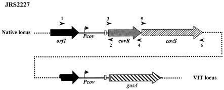
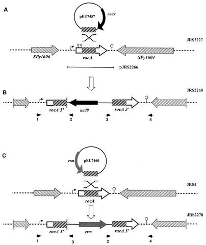
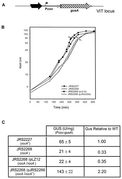
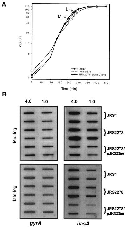
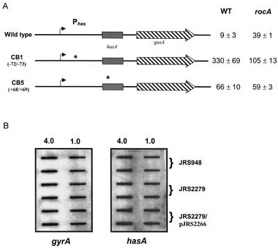
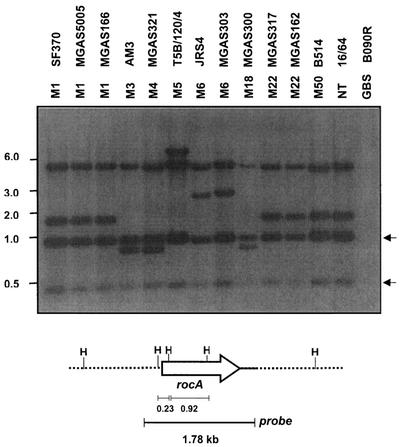
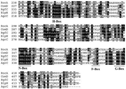
In the group A streptococcus (GAS; Streptococcus pyogenes), a two-component system known as CovRS (or CsrRS) regulates about 15% of the genes, including several important virulence factors like the hyaluronic acid capsule. Most of these genes, including covR itself, are negatively regulated by CovR. We have isolated two independent ISS1 insertions in an open reading frame (ORF) that increases CovR expression as measured by a Pcov-gusA reporter fusion in single copy in the GAS chromosome. This ORF, named rocA for "regulator of Cov," activates covR transcription about threefold. As expected, a rocA mutant is mucoid and produces more transcript from the has promoter since this promoter is repressed by CovR. This effect is dependent on the presence of a wild-type covR gene. In contrast to its activation of Pcov, RocA negatively regulates its own expression. This autoregulation is not dependent on the presence of the covR gene. All the phenotypes of the rocA mutant were complemented by the presence of the rocA gene on a plasmid. The rocA gene is present in strains of all nine M serotypes of GAS tested and is absent from strains representing 11 other groups of streptococci and related bacteria, including strains of the closely related group C and G streptococci. It seems likely that rocA plays an important role in the pathogenesis of GAS since it affects expression of the global regulator CovR.


Figures

FIG. 1.
FIG. 1.
Construction of Pcov-gusA reporter strain. The promoter region of covR, along with the sequence encoding the first two amino acids of CovR (amplified by primers 1 and 2 [arrowheads]), was fused to the gusA reporter gene isolated from plasmid pNZ8008. The Pcov-gusA reporter construct was inserted into the GAS chromosome at the VIT locus, which is not linked to the native covRS locus. Native covRS and the region of covR that is present at the VIT locus are shown. Symbols: bent arrow, covR promoter; white box, ribosome binding site of CovR; checkered box in front of gusA, first two amino acids of CovR; broken black arrow, part of the upstream ORF present in the Pcov-gusA fusion. Arrowheads indicate primers used for PCR analysis. The orientation of Pcov-gusA relative to the native covRS locus is unknown.
FIG. 2.
FIG. 2.
Construction of a rocA null mutant. (A) Region surrounding rocA in the chromosome of GAS. Plasmid pEU7457 (circle), which was used for insertional inactivation, contains a region internal to the rocA ORF (thick hatched box) and contains aad9, which encodes spectinomycin resistance (thick black arrow). The direction of transcription of the ORFs flanking rocA is indicated by striped arrowheads. The region of rocA that was cloned in complementing plasmid pJRS2266 is indicated by a bar below the chromosome. (B) JRS2268 was produced by homologous recombination (indicated by the X above the representation of the chromosome in panel A), which inserted pEU7457 into the wild-type rocA gene in the JRS2227 chromosome. (C) JRS2278 was produced by a similar targeted insertion in JRS4 by using integrational plasmid pEU7460, which contains erm, encoding erythromycin resistance (gray box), and a region internal to the rocA ORF (thick hatched box) as shown. Symbols: dotted lines, chromosome; striped boxes, coding regions with the directions of transcription indicated by the arrowheads; bent arrow, putative promoter; triangles, ISS1 insertion sites; lollipop, putative rho-independent transcription terminator. Small arrowheads below the chromosome represent primers used to confirm plasmid insertion into the chromosome (see Materials and Methods). The figure is not drawn to scale.
FIG. 3.
FIG. 3.
Expression of the covR gene in the wild-type and rocA mutant strains. (A) A gusA reporter system was used to measure the expression from the covR promoter at the VIT locus (symbols are as in Fig. 1). (B) Arrows indicate the times at which the samples were assayed for GusA activity. (C) The values shown are units of glucuronidase activity (with standard errors of the mean of experiments repeated at least four times). The relative ratio of GusA activity with respect to that of the wild type (WT) is shown.
FIG. 4.
FIG. 4.
Analysis of hasA gene transcription in the wild-type and rocA mutant strains. (A) Total RNA was isolated from JRS4 (wild type), JRS2278 (rocA), and JRS2278/pJRS2266 (rocA/rocA+) at the mid-exponential growth phase (M) and the late exponential growth phase (L). (B) On each filter, two dilutions (4.0 and 1.0 μg) of RNA were applied in vertically arranged duplicates. Membranes were probed with PCR-derived specific DNA probes internal to the coding region of gyrA and hasA (Materials and Methods). Results reported are representative of hybridization from three independent RNA isolations.
FIG. 5.
FIG. 5.
Regulation of the has promoter by rocA requires functional CovR. (A). Stars indicate mutations in the has promoter region that make it insensitive to CovR repression. The name of the mutant is on the left, and the location of the mutation is in parentheses. The values shown are glucuronidase units with standard errors of the mean of at least four independent experiments. WT, wild type. (B) Transcription analysis of hasA by RNA hybridization. RNA was isolated at the mid-exponential phase from JRS948 (covR), JRS2279 (covR rocA), and JRS2279/pJRS2266 (covR rocA/rocA+). Blots were prepared and hybridized as described in the legend to Fig. 4. Results are representative of hybridization from two independent RNA isolations.
FIG. 6.
FIG. 6.
RocA regulates its own expression. RNA was extracted from the JRS4 (wild-type) and JRS2278 (rocA) strains at the mid-exponential (ME) and late exponential (LE) phases as indicated in Fig. 4. Membranes were hybridized with gyrA and rocA probes. Blots were prepared and hybridized as described in Materials and Methods. The results shown are representative of hybridization from two independent RNA isolations.
FIG. 7.
FIG. 7.
Regulation of rocA expression by RocA is independent of functional CovR. RNA was extracted from the JRS4 (wild-type) and JRS948 (covR) strains at the late exponential (LE) and early stationary (ES) phases (arrows in panel A). Membranes were hybridized with rpsL, rocA, and sagA probes as described in Materials and Methods (B). The results shown are representative of hybridization from two independent RNA isolations.
FIG. 8.
FIG. 8.
Detection of the rocA gene by Southern hybridization. Genomic DNA from the strains was restricted with HindIII and separated in a 0.7% agarose gel by electrophoresis, and the presence of the rocA gene was detected by hybridization with a 1.78-kb fragment of the rocA region as shown at the bottom. M serotypes and strain numbers are indicated at the top. GBS, group B streptococcus; NT, nontypeable. Arrows indicate the constant fragments hybridized with the probe in all GAS strains. The values on the left are molecular sizes in kilobases.
FIG. 9.
FIG. 9.
Comparison of RocA with homologous proteins identified by a Blast-P search. The C-terminal regions of the different sensor kinases were aligned by using ClustalW. The GenBank accession numbers of the sequences are as follows: RocA, AAK34382; ComD, CAB39530; RgfC, AAM22582; BlpH, AAK74685; AgrC, AAB63269. Putative conserved boxes are indicated below the alignment.

References

    1. Alouf, J., and H. Muller-Alouf. 1996. Cellular constituents and extracellular proteins involved in the pathogenic capacity of Streptococcus group A. Ann. Pharm. Fr. 54:49-59. - PubMed
    1. Bernish, B., and I. van de Rijn. 1999. Characterization of a two-component system in Streptococcus pyogenes which is involved in regulation of hyaluronic acid production. J. Biol. Chem. 274:4786-4793. - PubMed
    1. Bisno, A. L. 1991. Group A streptococcal infections and acute rheumatic fever. N. Engl. J. Med. 325:783-793. - PubMed
    1. Biswas, I., P. Germon, K. McDade, and J. R. Scott. 2001. Generation and surface localization of intact M protein in Streptococcus pyogenes are dependent on sagA. Infect. Immun. 69:7029-7038. - PMC - PubMed
    1. Bronze, M. S., and J. B. Dale. 1996. The reemergence of serious group A streptococcal infections and acute rheumatic fever. Am. J. Med. Sci. 311:41-54. - PubMed

Publication types

Substances