. 2003 May;185(10):3147-54.
doi: 10.1128/JB.185.10.3147-3154.2003.

Early colonization events in the mutualistic association between Steinernema carpocapsae nematodes and Xenorhabdus nematophila bacteria

Affiliations

Early colonization events in the mutualistic association between Steinernema carpocapsae nematodes and Xenorhabdus nematophila bacteria

Eric C Martens et al. J Bacteriol. 2003 May.

Abstract

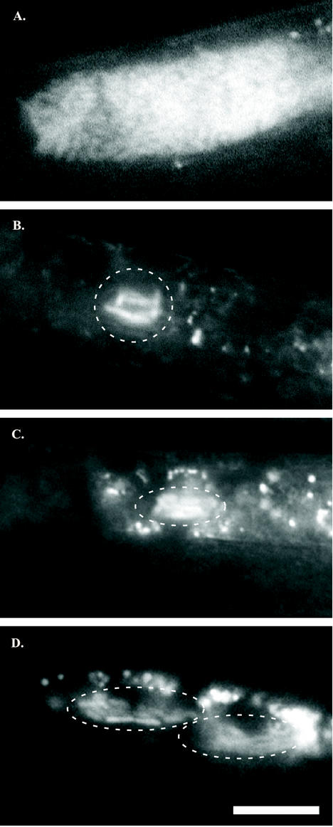
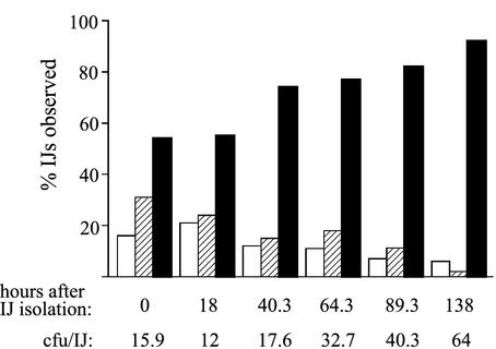
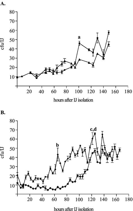
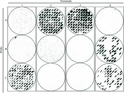
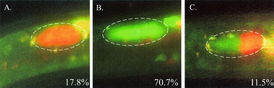
The bacterium Xenorhabdus nematophila is a mutualist of the entomopathogenic nematode Steinernema carpocapsae. During its life cycle, the bacterium exists both separately from the nematode and as an intestinal resident of a nonfeeding nematode form, the infective juvenile (IJ). The progression of X. nematophila from an ex vivo existence to a specific and persistent colonization of IJs is a model to understand the mechanisms mediating the initiation and maintenance of benign host-microbe interactions. To help characterize this process, we constructed an X. nematophila strain that constitutively expresses green fluorescent protein, which allowed its presence to be monitored within IJs. Using this strain, we showed that few bacterial cells initiate colonization of an individual IJ and that these grow inside the lumen of the IJ intestine in a reproducible polyphasic pattern during colonization. In accordance with these two observations, we demonstrated that the final population of bacteria in a nematode is of predominantly monoclonal origin, suggesting that only one or two bacterial clones initiate or persist during colonization of an individual nematode. These data suggest that X. nematophila initiates IJ colonization by competing for limited colonization sites or resources within the nematode intestine. This report represents the first description of the biological interactions occurring between X. nematophila and S. carpocapsae during the early stages of the colonization process, provides insights into the physiology of X. nematophila in its host niche, and will facilitate interpretation of future data regarding the molecular events mediating this process.


Figures

FIG. 1.
FIG. 1.
Bacterial colonization of immature IJs. Vesicles of mature IJs may contain 40 to >100 X. nematophila cells (A), whereas the vesicles of immature IJs contain only a few X. nematophila cells (indicated by enclosure in a dashed white line) (B to D). In panel B, six to seven rod-shaped X. nematophila cells cluster in close proximity to each other in the vesicle. The vesicle shown in panel C contains slightly more X. nematophila cells, and that in panel D contains even more than the IJ shown in panel B, but each has noticeably fewer than the full complement of cells found in a mature IJ (A). GFP-labeled X. nematophila cells were distinguished from nematode intestinal autofluorescence by virtue of bacterial cell shape and differential spectral emission under appropriate fluorescence filters (see Materials and Methods). In all images, nematodes are oriented with heads off the left side of the panel. Magnification, ×600. Bar, 10 μm.
FIG. 2.
FIG. 2.
Microscopic analysis of colonization density of GFP-labeled X. nematophila in nematode IJs. Nematodes were cultivated on lawns of HGB340, and immature IJs were isolated by repeated rinsing with sterile dH2O (see Materials and Methods). At various times after isolation (indicated below each set of bars), nematodes were observed by fluorescence microscopy and rated as belonging to one of three classes: not visibly colonized (open bars), oligocolonized (hatched bars), or fully colonized (filled bars). Between 170 and 303 IJs were observed for each time point, and at each time point a sample of IJs was used to quantify the average CFU per IJ (values shown below graph).
FIG. 3.
FIG. 3.
X. nematophila growth in isolated immature IJs. Populations of immature IJs were assayed at 4- or 6-h intervals to determine the average number of X. nematophila cells associated with IJs. Two replicates each from two separate experiments (A and B) are shown. Each point is the average result for three individual assays ± standard error (measured in CFU per IJ). Lowercase letters a to d indicate the regions of growth where data were used to determine the maximum growth rate (see Materials and Methods). Peaks from both curves under the region labeled c and d were used.
FIG. 4.
FIG. 4.
Competition of signature-tagged X. nematophila strains for nematode colonization. X. nematophila bacteria recovered from individual IJs (numbered 1 to 4) were probed to determine if they hybridized to probe A, B, or C. All 88 colonies recovered from IJ 1 hybridized to probe C. All 88 colonies recovered from IJs 2 and 3 hybridized to probe A. For IJ 4, 49% (43 of 88) of the clones hybridized to probe B, whereas the remaining 51% (45 of 88) hybridized to probe C.
FIG. 5.
FIG. 5.
Competition of HGB566 and HGB567 for nematode colonization. IJs raised on a lawn inoculated with equal amounts of HGB566 and HGB567 were analyzed to determine whether they contained only strain HGB567 (A), only strain HGB566 (B), or a mixture of both fluorescent strains (C). The frequency of each colonization type is indicated in the lower right corner of each panel. A total of 481 IJs were examined. The reason for the disproportionately high percentage of nematodes colonized by ECFP-expressing bacteria only may be due to the unequal competition of these strains for growth and survival during the ∼10-day coculture period. This inequality in strain representation further exemplifies the tendency toward monoclonal colonization because the 17.8% of nematodes that contained only DsRed-expressing bacteria would have been fully colonized by those bacteria despite the fact that they were a minority population.

References

    1. Aballay, A., and F. M. Ausubel. 2002. Caenorhabditis elegans as a host for the study of host-pathogen interactions. Curr. Opin. Microbiol. 5:97-101. - PubMed
    1. Akhurst, R. J. 1983. Neoaplectana species: specificity of association with bacteria of the genus Xenorhabdus. Exp. Parasitol. 55:258-263. - PubMed
    1. Akhurst, R. J., and N. Boemare. 1990. Biology and taxonomy of Xenorhabdus, p. 75-87. In R. Gaugler and H. K. Kaya (ed.), Entomopathogenic nematodes in biological control. CRC Press, Inc., Boca Raton, Fla.
    1. Ausubel, F. A., R. Brent, R. E. Kingston, D. D. Moore, J. G. Seidman, J. A. Smith, and K. Struhl. 1998. Current protocols in molecular biology. Wiley and Sons, New York, N.Y.
    1. Bao, Y., D. P. Lies, H. Fu, and G. P. Roberts. 1991. An improved Tn7-based system for the single-copy insertion of cloned genes into chromosomes of gram-negative bacteria. Gene 109:167-168. - PubMed

Publication types

LinkOut - more resources