

doi: 10.1128/JB.185.10.3228-3231.2003.
The Bacillus subtilis acyl lipid desaturase is a delta5 desaturase
Affiliations
- PMID: 12730185
- PMCID: PMC154086
- DOI: 10.1128/JB.185.10.3228-3231.2003
Item in Clipboard
The Bacillus subtilis acyl lipid desaturase is a delta5 desaturase
J Bacteriol.
2003 May.
Abstract
Bacillus subtilis was recently reported to synthesize unsaturated fatty acids (UFAs) with a double bond at positions delta5, delta7, and delta9 (M. H. Weber, W. Klein, L. Muller, U. M. Niess, and M. A. Marahiel, Mol. Microbiol. 39:1321-1329, 2001). Since this finding would have considerable importance in the double-bond positional specificity displayed by the B. subtilis acyl lipid desaturase, we have attempted to confirm this observation. We report that the double bond of UFAs synthesized by B. subtilis is located exclusively at the delta5 position, regardless of the growth temperature and the length chain of the fatty acids.
Figures
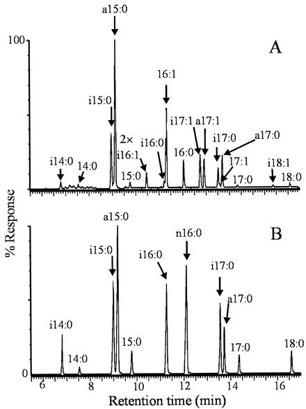
Chromatogram of fatty acids synthesized by strains AKP20 (A) and LC5 (B) at 25°C. The peaks corresponding to the identified fatty acids are indicated by arrows. i, iso-branched chain fatty acids; a, anteiso-branched chain fatty acids. The peaks of the chromatogram shown in panel A and located at the right of the 2× label were twofold magnified.
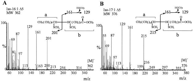
Mass spectrum of dimethyl disulfide derivatives separated by GC of iso-C16:1 (A) and iso-C17:1 (B). The aliphatic fragments a and b containing the carboxyl group are indicated by braces. [M]+, molecular ion.
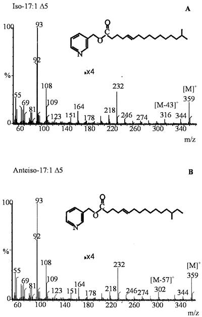
Mass spectra of 3-picolinyl esters. [M]+, molecular ion. The peaks of the mass spectra located at the right of the 4× label were fourfold magnified. The fragments at intervals of 43 and 57 amu ([M-43]+ and [M-57]+, respectively) are indicated. Details of interpretation of the spectrum are given in the text.
References
-
- Bligh, E. G., and W. J. Dyer. 1959. A rapid method of total lipid extraction and purification. Can. J. Biochem. Physiol. 31:911-917. - PubMed
-
- Christie, W. 1989. Gas chromatography and lipids. The Oily Press, Ayr, Scotland.
Publication types
MeSH terms
Substances
LinkOut - more resources
Full Text Sources
