. 2003 Jul 4;278(27):24577-85.
doi: 10.1074/jbc.M304530200. Epub 2003 Apr 30.

Fibronectin fragment activation of proline-rich tyrosine kinase PYK2 mediates integrin signals regulating collagenase-3 expression by human chondrocytes through a protein kinase C-dependent pathway

Affiliations

Fibronectin fragment activation of proline-rich tyrosine kinase PYK2 mediates integrin signals regulating collagenase-3 expression by human chondrocytes through a protein kinase C-dependent pathway

Richard F Loeser et al. J Biol Chem. .

Abstract

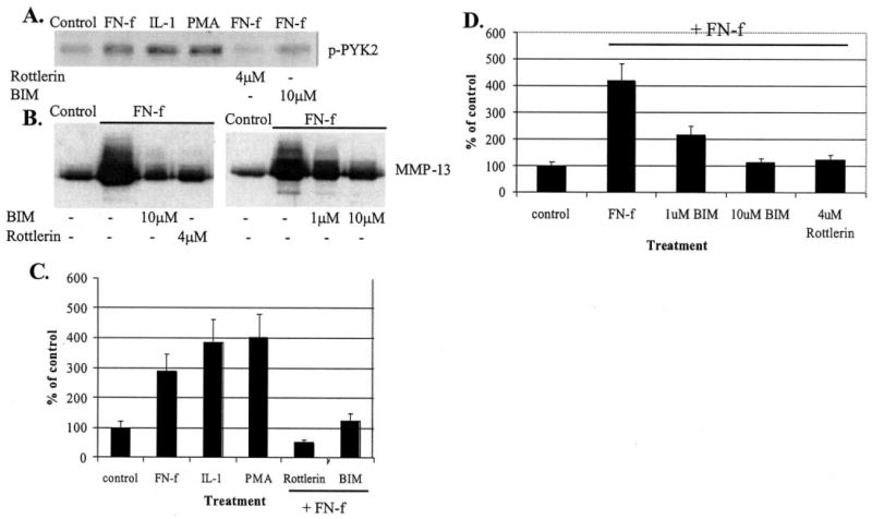
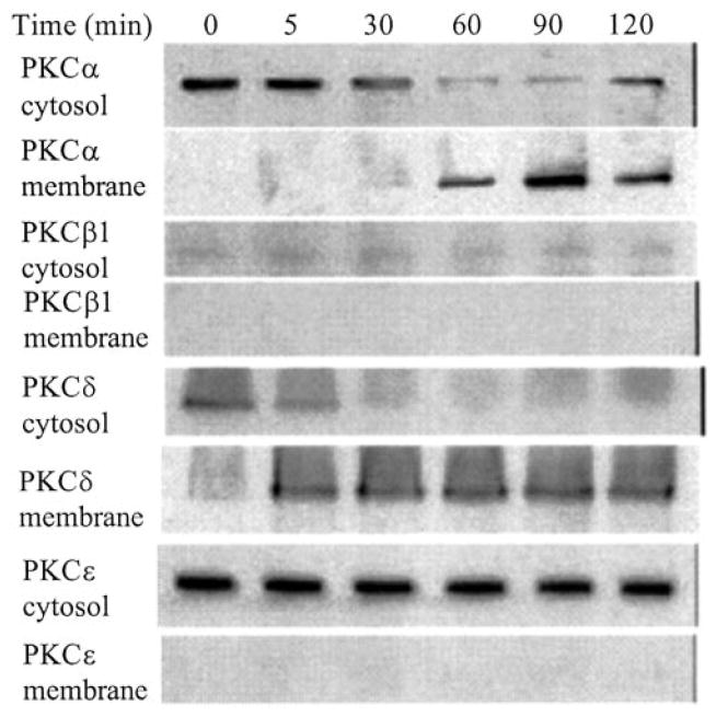
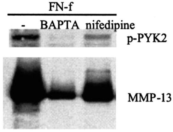
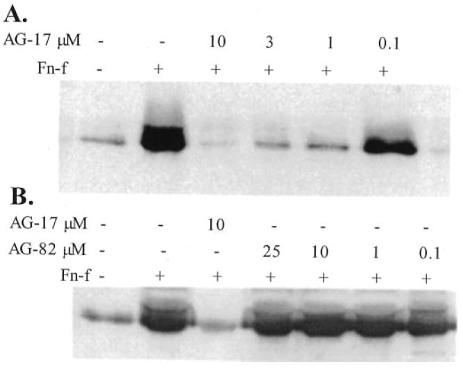
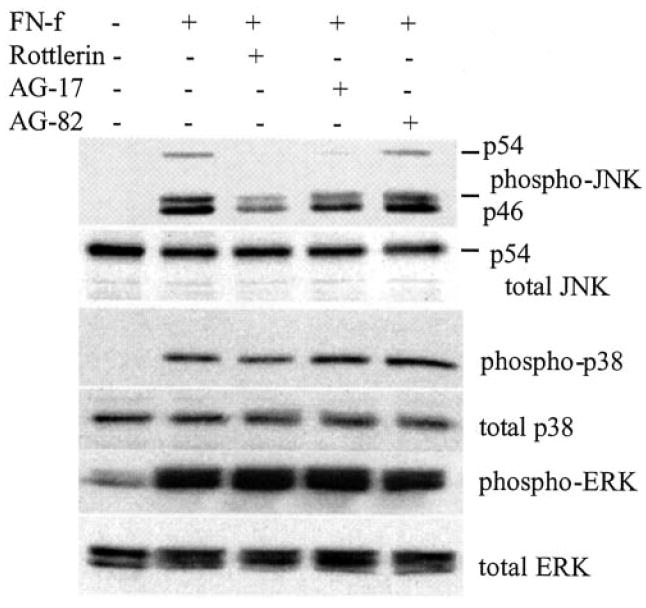
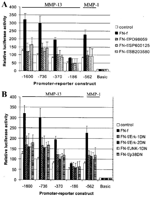
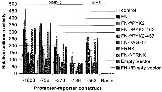
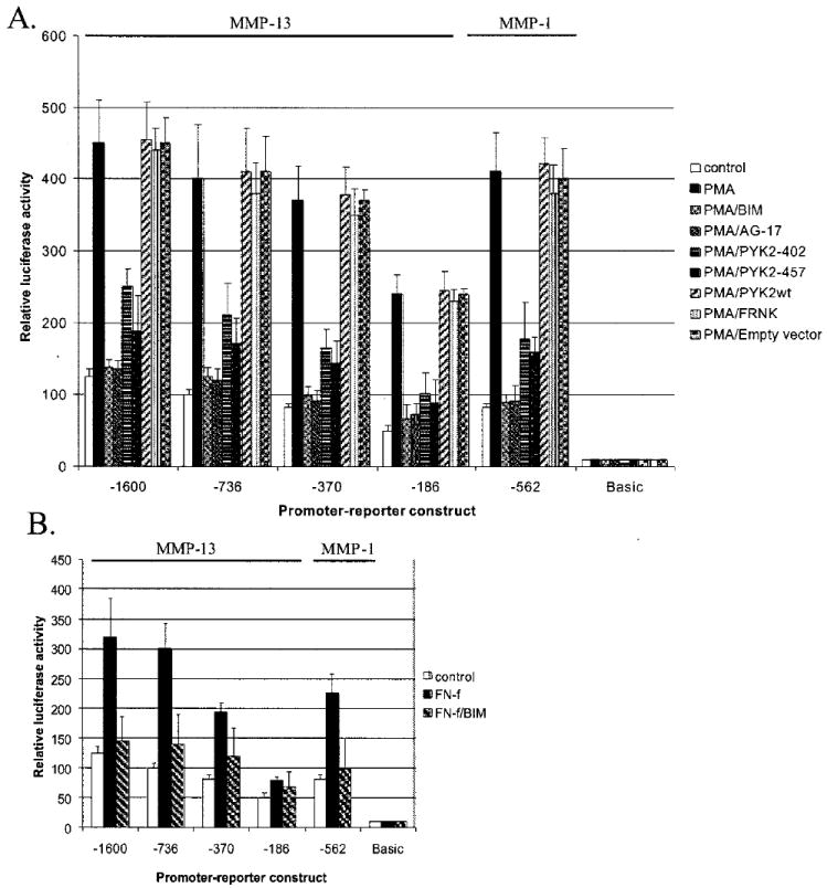
Fibronectin fragments (FN-f), including the 110-kDa fragment that binds the alpha5beta1 integrin, stimulate collagenase-3 (MMP-13) production and cartilage destruction. In the present study, treatment of chondrocytes with the 110-kDa FN-f or an activating antibody to the alpha5beta1 integrin was found to increase tyrosine autophosphorylation (Tyr-402) of the proline-rich tyrosine kinase-2 (PYK2) without significant change in autophosphorylation (Tyr-397) of focal adhesion kinase (FAK). The tyrosine kinase inhibitor tyrphostin A9, shown previously to block a PYK2-dependent pathway, blocked the FN-f-stimulated increase in MMP-13, whereas tyrphostin A25 did not. FN-f-stimulated PYK2 phosphorylation and MMP-13 production was also blocked by reducing intracellular calcium levels. Adenovirally mediated overexpression of wild type but not mutant PYK2 resulted in increased MMP-13 production. The protein kinase C (PKC) activator phorbol 12-myristate 13-acetate stimulated PYK2 phosphorylation and MMP-13 production. MMP-13 expression stimulated by either phorbol 12-myristate 13-acetate or FN-f was blocked by PKC inhibitors including the PKCdelta inhibitor rottlerin. Furthermore, PKCdelta translocation from cytosol to membrane was noted within 5 min of stimulation with FN-f. Immortalized human chondrocytes, transiently transfected with MMP-13 promoter-luciferase reporter constructs, showed increased promoter activity after FN-f treatment that was inhibited by co-transfection with either of two dominant negative mutants of PYK2 (Y402F and K457A). No inhibition was seen after cotransfection with wild type PYK2, a dominant negative of FAK (FRNK) or empty vector plasmid. FN-f-stimulated MMP-13 promoter activity was also inhibited by chemical inhibitors of ERK, JNK, and p38 mitogen-activated protein (MAP) kinases or by co-transfection of dominant negative MAP kinase mutant constructs. These studies have identified a novel pathway for the MAP kinase regulation of MMP-13 production which involves FN-f stimulation of the alpha5beta1 integrin and activation of the nonreceptor tyrosine kinase PYK2 by PKC, most likely PKCdelta


Figures

Fig. 1
Fig. 1. Phosphorylation of PYK2 in chondrocytes treated with fibronectin fragments or an α5β1 integrin antibody
Human articular chondrocyte cultures were made serum free and then stimulated with 1 μm of the 110-kDa FN-f (A) or 25 μg/ml of an activating antibody to the α5β1 integrin (B). Cell lysates were prepared at the indicated time points and used for immunoblotting with antibodies to the autophosphorylation sites of PYK (Tyr-402, p-PYK2) and FAK (Tyr-397, p-FAK). C, mean band intensity from immunoblots of cells treated with FN-f was calculated as % of time 0 controls for p-PYK2 (filled bars) and p-FAK (open bars).
Fig. 2
Fig. 2. PKC inhibition blocks fibronectin fragment stimulation of PYK2 phosphorylation and MMP-13 expression
A, human articular chondrocytes were treated for 30 min with control media or media containing 1 μm FN-f, 10 ng/ml IL-β, 50 nm PMA, or FN-f after pretreatment with 4 μm rottlerin or 10 μm BIM. Cell lysates were immunoblotted with antibodies to phosphorylated PYK2 (p-PYK2). B, conditioned media were collected from human articular chondrocytes treated for 24 h with FN-f with or without the indicated concentrations of BIM or rottlerin. Media samples were concentrated and equal amounts used for immunoblotting with antibodies to MMP-13. C and D, mean band intensity results for the experiments shown in A and B, respectively, expressed as % of unstimulated controls.
Fig. 3
Fig. 3. Chondrocyte PKC activation in response to fibronectin fragments
Human articular chondrocytes were stimulated for the indicated periods with 1 μm FN-f. Cytosol and membrane fractions were prepared, and samples equalized for total protein were immunoblotted with antibodies to the specific PKC isoforms.
Fig. 4
Fig. 4. Inhibition of intracellular calcium signaling blocks fibronectin fragment stimulation of PYK2 phosphorylation and MMP-13 expression
Human articular chondrocytes were pretreated for 30 min with control media or either 10 μm BAPTA-AM or 20 μm nifedipine and then stimulated with 1 μm FN-f. After 30 min, cell lysates were prepared and used for immunoblotting with antibodies to phosphorylated PYK2 (p-PYK2). Parallel cultures were allowed to incubate with FN-f and inhibitors for 24 h, and conditioned media were collected. Samples of equal volume were immunoblotted with antibodies to MMP-13.
Fig. 5
Fig. 5. Tyrosine kinase inhibition with tyrphostin A9 (AG-17) blocks fibronectin fragment stimulation of MMP-13 synthesis
Human articular chondrocytes were pretreated for 30 min with the increasing concentrations of tyrphostin A9 (AG-17) (A) or tyrphostin A25 (AG-82) (B) and then treated overnight with 1 μm FN-f or control media without FN-f. Conditioned media were used for immunoblotting with antibodies to MMP-13.
Fig. 6
Fig. 6. Inhibition of fibronectin fragment induced JNK phosphorylation by tyrphostin A9 (AG-17) and rottlerin
Human articular chondrocytes were pretreated for 30 min with the indicated inhibitors: 4 μm rottlerin, 10 μm tyrphostin A9 (AG-17), or 10 μm tyrphostin A25 (AG-82). The cells were then treated with 1 μm FN-f or control media without FN-f or inhibitors for 30 min at which time cell lysates were prepared. Chondrocyte lysates were immunoblotted with phosphospecific (phospho-) and non-phosphospecific (total) antibodies to JNK, p38, and ERK.
Fig. 7
Fig. 7. Adenoviral overexpression of PYK2 stimulates chondrocyte MMP-13 production
Primary chondrocytes were treated with adenovirus containing a GFP expression construct (GFP) as control or GFP linked to wild type PYK2 (wtPYK2), a kinase dead PYK2 (kd-PYK2), or Y402F-PYK2. A, cell lysates were immunoblotted with phosphospecific and non-phosphospecific (total) antibodies to PYK2. A sample of lysate from an untreated control culture was included (cntl). B, media samples from duplicate wells of adenoviral treated cells were analyzed for MMP-13 by immunoblotting. The positive control (+cntl) was from cells treated with FN-f without adenoviral treatment, whereas none of the adenoviral treated cultures had received FN-f.
Fig. 8
Fig. 8. MAP kinase inhibition blocks fibronectin fragment stimulation of MMP-13 and MMP-1 promoter activity
A, immortalized human chondrocytes (C-28I2 cells) were transiently transfected with −1600, −736, −370, −186 MMP-13, and −562 MMP-1 promoter-luciferase reporter constructs or the promoterless luciferase vector (basic) as control. After 24 h in complete media, the cells were changed to serum-free conditions for 20–24 h and then treated with 1 μm FN-f with or without MAP kinase inhibitors (30 μm PD98059, 20 μm SP600125, or 10 μm SB203580). After 24 h of incubation, cell lysates were prepared and used for luciferase assays. B, C-28I2 cells were co-transfected with the MMP promoter-reporter constructs and dominant negative (DN) mutant constructs for ERK-1, ERK-2, JNK, or p38 as indicated. Cells were incubated for 24 h in complete media, made serum-free for 24 h, and then treated with 1 μm FN-f for 24 h followed by analysis of luciferase activity.
Fig. 9
Fig. 9. Inhibition of PYK2 blocks fibronectin fragment stimulation of MMP-13 and MMP-1 promoter activity
Immortalized human chondrocytes were co-transfected with the MMP promoter-reporter constructs described in FIG. 8 and constructs for wild type PYK2, dominant negative PYK2 mutants (PYK2–402 and -457), dominant negative FAK (FRNK), or an empty control vector. Tyrosine kinase inhibition by 10 μm tyrphostin A9 (AG-17) was also tested. Following 24 h of incubation in complete media and 24 h incubation in serum-free media, the cultures were treated with 1 μm FN-f as indicated. After 24 h of treatment, cell lysates were prepared and analyzed by luciferase assay.
Fig. 10
Fig. 10. Inhibition of PYK2 blocks PMA-stimulated MMP-13 and MMP-1 promoter activity and PKC inhibition blocks fibronectin fragment stimulated activity
A, immortalized human chondrocytes were transiently transfected with MMP-13 and MMP-1 promoter-reporter constructs and in some cultures co-transfected with the PYK2 and FAK constructs as described in Fig. 9. The cultures were stimulated with 50 nM PMA with or without 5 μm BIM or 10 μm tyrphostin A9 (AG-17). After 24 h, cell lysates were prepared and used to measure luciferase activity. B, chondrocytes transiently transfected with the promoter-reporter constructs were treated with the FN-f in the absence or presence of 5 μm BIM.

References

    1. Sternlicht MD, Werb Z. Annu Rev Cell Dev Biol. 2001;17:463–516. - PMC - PubMed
    1. Dean DD, Martel-Pelletier J, Pelletier JP, Howell DS, Woessner JF., Jr J Clin Invest. 1989;84:678–685. - PMC - PubMed
    1. Billinghurst RC, Dahlberg L, Ionescu M, Reiner A, Bourne R, Rorabeck C, Mitchell P, Hambor J, Diekmann O, Tschesche H, Chen J, Van Wart H, Poole AR. J Clin Invest. 1997;99:1534–1545. - PMC - PubMed
    1. Shlopov BV, Lie WR, Mainardi CL, Cole AA, Chubinskaya S, Hasty KA. Arthritis Rheum. 1997;40:2065–2074. - PubMed
    1. Reboul P, Pelletier JP, Tardif G, Cloutier JM, Martel-Pelletier J. J Clin Invest. 1996;97:2011–2019. - PMC - PubMed

Publication types