. 2003 May 13;100(10):5736-41.
doi: 10.1073/pnas.1037393100. Epub 2003 May 2.

Tuning activation of the AMPA-sensitive GluR2 ion channel by genetic adjustment of agonist-induced conformational changes

Affiliations

Tuning activation of the AMPA-sensitive GluR2 ion channel by genetic adjustment of agonist-induced conformational changes

Neali Armstrong et al. Proc Natl Acad Sci U S A. .

Abstract

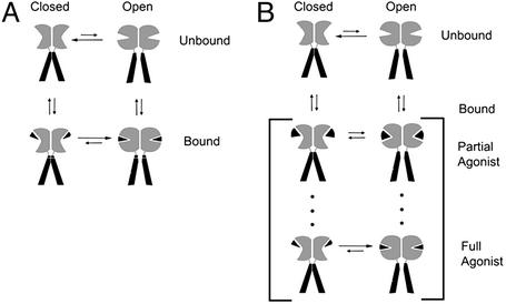
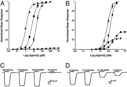
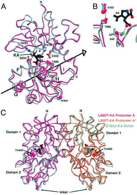
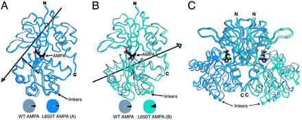
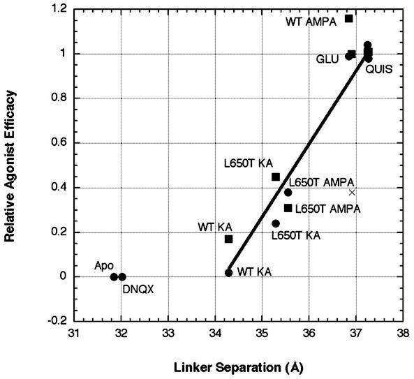
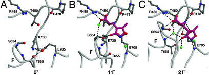
The (S)-2-amino-3-(3-hydroxy-5-methyl-4-isoxazole) propionic acid (AMPA) receptor discriminates between agonists in terms of binding and channel gating; AMPA is a high-affinity full agonist, whereas kainate is a low-affinity partial agonist. Although there is extensive literature on the functional characterization of partial agonist activity in ion channels, structure-based mechanisms are scarce. Here we investigate the role of Leu-650, a binding cleft residue conserved among AMPA receptors, in maintaining agonist specificity and regulating agonist binding and channel gating by using physiological, x-ray crystallographic, and biochemical techniques. Changing Leu-650 to Thr yields a receptor that responds more potently and efficaciously to kainate and less potently and efficaciously to AMPA relative to the WT receptor. Crystal structures of the Leu-650 to Thr mutant reveal an increase in domain closure in the kainate-bound state and a partially closed and a fully closed conformation in the AMPA-bound form. Our results indicate that agonists can induce a range of conformations in the GluR2 ligand-binding core and that domain closure is directly correlated to channel activation. The partially closed, AMPA-bound conformation of the L650T mutant likely captures the structure of an agonist-bound, inactive state of the receptor. Together with previously solved structures, we have determined a mechanism of agonist binding and subsequent conformational rearrangements.


Figures

Figure 1
Figure 1
Mechanisms to describe the conformational behavior of ligand-gated ion channels. (A) The two-state model where the receptor is in equilibrium between two conformations, closed and open. Agonist binding stabilizes the receptor in the open state. Full agonists stabilize the open state more effectively than partial agonists, and both types of agonists stabilize the same conformational states. (B) Multistate model where the agonist-binding region of the receptor adopts a range of agonist-dependent conformations. Partial agonists promote a submaximal conformational change and therefore are not as effective in shifting the closed to open equilibrium of the ion channel to the open state.
Figure 2
Figure 2
Dose–response curves and Imax traces for the L483Y and L483Y/L650T variants of the full-length GluR2 receptor recorded by using the two-electrode, voltage–clamp technique. (A) Normalized dose–response curves for glutamate (Glu) (⧫), AMPA (■), quisqualate (Quis) (▴), and kainate (KA) (●) measured from oocytes expressing GluR2 L483Y receptors. (B) EC50 data for the GluR2 L483Y/L650T mutant, in combination with glutamate (⧫), AMPA (■), quisqualate (▴), or kainate (●), scaled for efficacy relative to glutamate. (C) Maximal currents (Imax) elicited by L483Y receptors and saturating concentrations of agonist. The duration of agonist application is indicated by horizontal, thick black lines, and the identity and concentration of the agonist is indicated above each line. (D) Maximum currents mediated by L483Y/L650T receptors. The Imax measurements were made on five individual oocytes. All values are summarized in Table 1.
Figure 3
Figure 3
Comparison of the WT and S1S2J L650T/kainate conformations. (A) Superimposition of WT and L650T/kainate structures. The WT backbone is drawn in gray and the mutant structure is shown in magenta. Kainate is drawn in black. Selected binding cleft side chains are drawn in ball-and-stick representation and colored as the backbone. The large spheres at the bottom of the structure depict the location of the first residue (Gly) in the two residue Gly-Thr linker. (B) Close-up view of superimposed WT and mutant kainate (KA) binding sites. The green sphere is a water in the mutant structure and the purple sphere is a water in the WT structure. In the mutant structure, the hydroxyl of Thr-650 makes a water-mediated hydrogen bond with kainate that is absent in the WT structure. In the L650T/kainate structure, two key residues situated on the clamshell cleft move 0.6 Å closer together relative to the WT kainate structure, placing the hydroxyl of Thr-686 in ideal hydrogen bonding distance (2.6 Å) to a carboxylate oxygen of Glu-402. (C) Superimposed S1S2J/kainate and L650T/kainate crystallographic dimers. WT protomers are drawn in gray and mutant protomers are shown in magenta (A) and orange (A*-symmetry related protomer). Thr-650 is drawn in space-filling mode.
Figure 4
Figure 4
Comparison of WT and S1S2J L650T/AMPA(AS) conformations. (A) Superposition of WT S1S2J/AMPA (gray) with S1S2J L650T/AMPA (AS form) protomer A (blue). (B) Superposition of WT S1S2J/AMPA (gray) with S1S2J L650T/AMPA(AS) protomer B (green). The black arrows in A and B indicate the axis of rotation relating the conformational difference between the WT and L650T structures. (C) Superimposed WT and mutant AMPA dimers.
Figure 5
Figure 5
Correlation between linker separation and agonist efficacy. The values for relative agonist efficacy were determined for L483Y receptors (●) and WT receptors in the presence of 100 μM cyclothiazide (■) and are listed in Table 1. Apo and 6,7-dinitroquinoxaline-2,3-dione (DNQX) are assumed to have an efficacy of zero, and the other values are scaled to glutamate, which is set to one. Linker separation is defined as the distance between Gly Cα atoms from the artificial Gly-Thr linker joining S1 and S2. The structure corresponding to each point is indicated. The L650T/AMPA(Zn) structure is indicated with an ×. The glutamate (Glu) and quisqualate (Quis) points are all clustered around an efficacy of 1.0. The agonist-bound data points were fit with a linear equation that yields a correlation coefficient of 0.893. KA, kainate.
Figure 6
Figure 6
Mechanism of agonist binding and domain closure. (A) The binding site of the open-cleft, closed-channel state (Apo S1S2J, protomer A). (B) The possible first step in agonist binding as observed in molecule B of the L650T/AMPA(AS) structure. We suggest that this semiclosed cleft conformation represents the agonist-bound, closed-channel state. (C) The closed-cleft, open-channel conformation as observed in the WT S1S2J/AMPA binding cleft (protomer A). In B and C, water molecules are shown as green spheres, AMPA is drawn in magenta, and hydrogen bonds are depicted by black dashed lines. The degrees of domain closure relative to the Apo conformation are indicated below each structure.

References

    1. Hille B. Ion Channels of Excitable Membranes. Sunderland, MA: Sinauer; 2001.
    1. Monod J, Wyman J, Changeux J P. J Mol Biol. 1965;12:88–118. - PubMed
    1. Koshland D E. Proc Natl Acad Sci USA. 1958;44:98–104. - PMC - PubMed
    1. Koshland D E, Nemethy G, Filmer D. Biochemistry. 1966;5:365–385. - PubMed
    1. Li J, Zagotta W N, Lester H A. Q Rev Biophys. 1997;30:177–193. - PubMed

Publication types

MeSH terms

Substances

LinkOut - more resources