. 2003 May 13;100(10):6033-8.
doi: 10.1073/pnas.1135965100. Epub 2003 May 2.

Deletion of neuropeptide Y (NPY) 2 receptor in mice results in blockage of NPY-induced angiogenesis and delayed wound healing

Affiliations

Deletion of neuropeptide Y (NPY) 2 receptor in mice results in blockage of NPY-induced angiogenesis and delayed wound healing

A Jonas Ekstrand et al. Proc Natl Acad Sci U S A. .

Erratum in

  • Proc Natl Acad Sci U S A. 2007 Feb 13;104(7):2554

Abstract

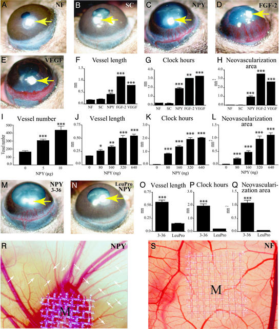
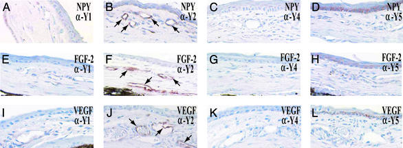
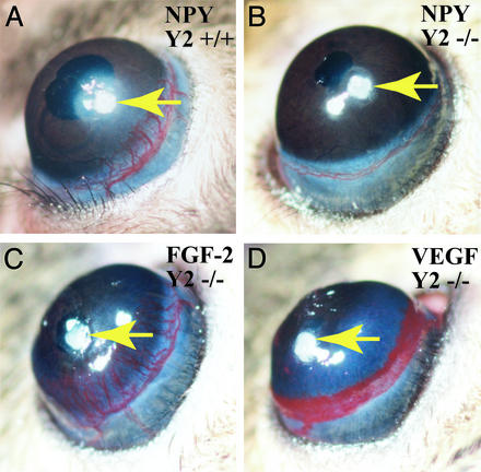
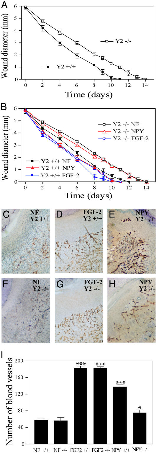
Neuropeptide Y (NPY), a 36-aa peptide, is widely distributed in the brain and peripheral tissues. Whereas physiological roles of NPY as a hormoneneurotransmitter have been well studied, little is known about its other peripheral functions. Here, we report that NPY acts as a potent angiogenic factor in vivo using the mouse corneal micropocket and the chick chorioallantoic membrane (CAM) assays. Unlike vascular endothelial growth factor (VEGF), microvessels induced by NPY had distinct vascular tree-like structures showing vasodilation. This angiogenic pattern was similar to that induced by fibroblast growth factor-2, and the angiogenic response was dose-dependent. In the developing chick embryo, NPY stimulated vascular sprouting from preexisting blood vessels. When [Leu(31)Pro(34)]NPY, a NPY-based analogue lacking high affinity for the NPY Y(2) receptor but capable of stimulating both Y(1) and Y(5) receptors, was used in the corneal model, no angiogenic response could be detected. In addition, NPY failed to induce angiogenesis in Y(2) receptor-null mice, suggesting that this NPY receptor subtype was mediating the angiogenic signal. In support of this finding, the Y(2) receptor, but not Y(1), Y(4), or Y(5) receptors, was found to be widely expressed in newly formed blood vessels. Further, a delay of skin wound healing with reduced neovascularization was found in Y(2) receptor-null mice. These data demonstrate that NPY may play an important role in the regulation of angiogenesis and angiogenesis-dependent tissue repair.


Figures

Figure 1
Figure 1
In vivo angiogenic activity of NPY. Micropellets containing 160 ng of NPY (C), 80 ng of FGF-2 (D), or 160 ng of VEGF (E) were implanted into corneal micropockets of C57BL/6 mice as described in Materials and Methods. Slow-release polymers containing no factors (A, NF) or 160 ng of a scrambled peptide (B, SC) served as negative controls. Corneal neovascularization was measured and photographed with a slit-lamp stereomicroscope on day 5 after growth factor implantation. Arrows point to the implanted pellets. Photographs represent ×20 amplification of the mouse eye. Quantitation of corneal neovascularization is presented as maximal vessel length (F), clock hours of circumferential neovascularization (G), and area of neovascularization (H). Graphs represent mean values (±SEM) of 10 eyes (five to seven mice) in each group. Various amounts (80, 160, 320, and 640 ng) of NPY (JL) were implanted into the micropockets of mouse corneas. Corneal neovascularization was measured as vessel length (J), clock-hours (K), and area (L) with a slit-lamp stereomicroscope on day 5 after growth factor implantation. Graphs represent mean values (±SEM) of 10 eyes (five to seven mice) in each group. Micropellets containing an equal amount (160 ng per pellet) of NPY3–36 (M) or [Leu31Pro34]NPY (N) were implanted into the corneas of C57BL/6 mice. The corneal neovascularization was examined and photographed on day 5 after pellet implantation. Arrows in M and N indicate the implanted pellet. Angiogenic responses were measured as vessel length (O), clock-hours of neovascularization (P), and vascular area (Q). Graphs represent mean values (±SEM) of 11–16 eyes (six to eight mice) in each group. Nylon meshes, M (9.3 mm2), coated with 0.45% methylcellulose containing various amounts of NPY or BSA were implanted on CAMs of 9-day-old chick embryos. After 5-day implantation, the formation of new blood vessels was examined under a stereomicroscope. A CAM with a methylcellulose mesh containing BSA (NF) alone served as a negative control (S). R shows an example of 5 μg of NPY-implanted CAM. New blood vessels are marked with arrows in R. The average of total numbers of microvessels within a defined area of 25 mm2 surrounding the implanted mesh is presented (I). *, P < 0.05; **, P < 0.01; ***, P < 0.001.
Figure 2
Figure 2
Localization of NPY Y2 receptor on the newly formed blood vessels. Histological sections of NPY-implanted (AD), FGF-2-implanted (EH), and VEGF-implanted (IL) corneas were incubated with an anti-Y1 (A, E, and I), an anti-Y2 (B, F, and J), an anti-Y4 (C, G, and K), or an anti-Y5 (D, H, and L) receptor antibody and stained with a peroxidase-conjugated secondary antibody. Endothelial cells of corneal microvessels were immunoreactive as indicated by arrows (B, F, and J).
Figure 3
Figure 3
Absence of corneal neovascularization in Y2 receptor null mice. NPY at the amount of 160 ng per pellet was implanted into each cornea of NPY Y2 +/+ (A) and NPY Y2−/− (B) mice. As controls, 80 ng per pellet of FGF-2 (C) or 160 ng per pellet of VEGF (D) was implanted into each of NPY Y2−/− mice. After 5-day implantation, corneal neovascularization was detected and pictured. Six mice and 12 corneas were used in each group of mice.
Figure 4
Figure 4
Delay of skin wound healing in Y2 receptor-deficient mice. Full-thickness skin wounds (6 mm in diameter) were created on the back of shaved NPY Y2+/+ and NPY Y2−/− mice by using a template. In B, each group of animals (six to seven per group) was topically treated with slow-release polymers containing NPY, FGF-2, or PBS (NF) as indicated. Diameters of wounds were measured daily (A and B). Data are presented in A and B as mean determinants (±SEM) of wounds of six to seven mice in each group. At day 7 after implantation, some wounds in each group were removed for immunohistochemical analysis by using an anti-CD31 staining antibody. Positive immunostaining signals of blood vessels in wounds of NPY Y2+/+ mice (CE) and of Y2−/− mice (FH) treated with PBS (C and F), FGF-2 (D and G), and NPY (E and H) were revealed by a peroxidase reaction. NF, no factor (PBS). Vascular density was quantified by counting microvessel numbers in six random fields under ×40 magnification (I). The average numbers of vessels (±SEM) are presented.

Similar articles

Cited by

References

    1. Blomqvist A G, Herzog H. Trends Neurosci. 1997;20:294–298. - PubMed
    1. Larhammar D. Regul Pept. 1996;65:165–174. - PubMed
    1. Franco-Cereceda A, Lundberg J M, Dahlof C. Acta Physiol Scand. 1985;124:361–369. - PubMed
    1. Erlinge D, Brunkwall J, Edvinsson L. Regul Pept. 1994;50:259–265. - PubMed
    1. Zukowska-Grojec Z, Pruszczyk P, Colton C, Yao J, Shen G H, Myers A K, Wahlestedt C. Peptides. 1993;14:263–268. - PubMed

Publication types