. 2003 May 12;161(3):497-505.
doi: 10.1083/jcb.200303030. Epub 2003 May 5.

Oscillatory nucleocytoplasmic shuttling of the general stress response transcriptional activators Msn2 and Msn4 in Saccharomyces cerevisiae

Affiliations

Oscillatory nucleocytoplasmic shuttling of the general stress response transcriptional activators Msn2 and Msn4 in Saccharomyces cerevisiae

Michel Jacquet et al. J Cell Biol. .

Abstract

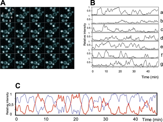
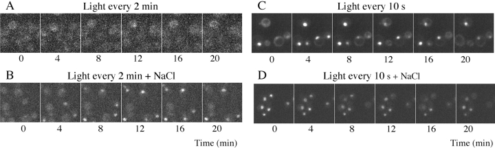
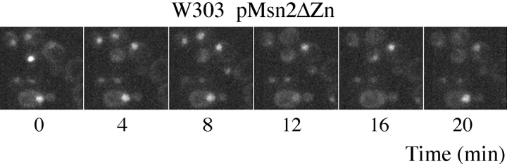
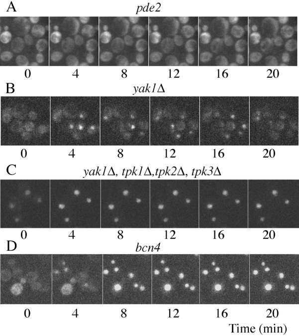
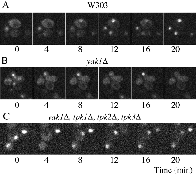
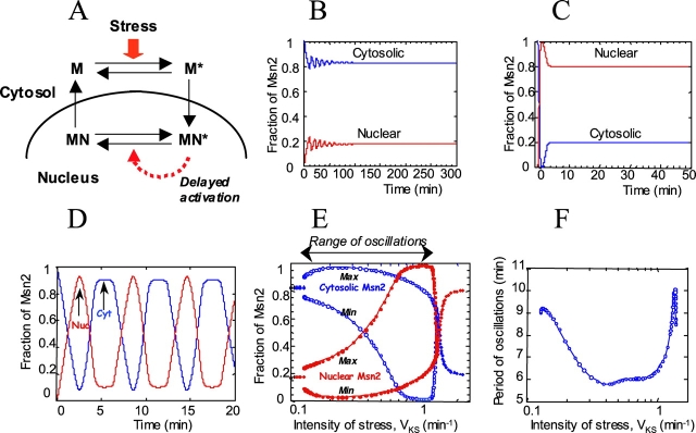
Msn2 and Msn4 are two related transcriptional activators that mediate a general response to stress in yeast Saccharomyces cerevisiae by eliciting the expression of specific sets of genes. In response to stress or nutritional limitation, Msn2 and Msn4 migrate from the cytoplasm to the nucleus. Using GFP-tagged constructs and high-resolution time-lapse video microscopy on single cells, we show that light emitted by the microscope also triggers this migration. Unexpectedly, the population of Msn2 or Msn4 molecules shuttles repetitively into and out of the nucleus with a periodicity of a few minutes. A large heterogeneity in the oscillatory response to stress is observed between individual cells. This periodic behavior, which can be induced by various types of stress, at intermediate stress levels, is not dependent upon protein synthesis and persists when the DNA-binding domain of Msn2 is removed. The cAMP-PKA pathway controls the sensitivity of the oscillatory nucleocytoplasmic shuttling. In the absence of PKA, Msn4 continues to oscillate while Msn2 is maintained in the nucleus. We show that a computational model based on the possibility that Msn2 and Msn4 participate in autoregulatory loops controlling their subcellular localization can account for the oscillatory behavior of the two transcription factors.


Figures

Figure 1.
Figure 1.
Oscillatory nucleocytoplasmic shuttling of Msn2. Video microscopic examination of W303 yeast cells containing the plasmid pAMG growing in YNB glucose at 30°C. The subcellular localization of Msn2–GFP was determined by means of time-lapse video microscopy. Fluorescent images were recorded over 48 min. (A) A sequence of pictures taken every minute during the first 20 min, displayed from left to right and top to bottom. (B) The normalized variation of fluorescence in the nucleus of the cells labeled a–g in A. (C) The kinetics of nuclear (red line) and cytoplasmic (blue line) intensity of fluorescence for cell h in A. Curves presented in B and C have been drawn from values taken every 10 s and smoothened by taking the average between two consecutive values (see Video 1, available at http://www.jcb.org/cgi/content/full/jcb.200303030/DC1).
Figure 2.
Figure 2.
Effect of light and stresses on Msn2 subcellular localization. The same cells as in Fig. 1 were examined under various conditions of illumination as indicated above the images. (A and B) Pictures were taken every 2 min with a light intensity reduced to a minimum, giving a fourfold reduction in GFP fluorescence. No other stress was applied in A, whereas 0.5 M NaCl was added to the cell prior to examination in B. (C and D) Standard illumination was used (light applied for 1 s every 10 s) without other stress (C) and with 0.5 M NaCl (D). A subset of images taken at 4-min intervals is presented.
Figure 3.
Figure 3.
Oscillatory behavior of Msn2ΔZn–GFP. Video-microscopic images of localization are presented for a W303 cell containing pMsn2ΔZn–GFP (see Video 2, available at http://www.jcb.org/cgi/content/full/jcb.200303030/DC1).
Figure 4.
Figure 4.
Effect of mutations in the cAMP–PKA system. Time-lapse video microscopy images of Msn2–GFP localization in various mutants of the cAMP–PKA system are presented. pde2 stands for the OL564-1B strain with the phosphodiesterase mutation rca1 in the PDE2 gene (Wilson et al., 1993). yak1Δ corresponds to the OL607-47B strain deleted for this protein kinase. yak1Δ, tpk1Δ, tpk2Δ, tpk3Δ corresponds to the OL625-3 strain lacking all three PKA catalytic subunits (see Video 3, available at http://www.jcb.org/cgi/content/full/jcb.200303030/DC1). bcn4 corresponds to the strain BC-N4 in which the BCY1 gene coding for the regulatory subunit of PKA has been deleted from the NH2-terminal part responsible for PKA nuclear localization (Griffioen et al., 2001).
Figure 5.
Figure 5.
Oscillatory behavior of Msn4–GFP. Msn4–GFP behavior is shown in W303 cells (A), in yak1Δ cells (B), and in yak1Δ, tpk1Δ, tpk2Δ, tpk3Δ cells (C). Only the images taken at 4-min intervals are presented (see Video 3, available at http://www.jcb.org/cgi/content/full/jcb.200303030/DC1).
Figure 6.
Figure 6.
Computational model for oscillatory nucleocytoplasmic shuttling of the transcription factor Msn2. (A) A schematic model showing interconversions between the different states of Msn2: cytoplasmic, inactive (M); cytoplasmic, primed to enter the nucleus (M*); nuclear, active (MN*); nuclear, primed to leave the nucleus (MN). In the absence of stress, form M predominates. Under stress, the nuclear form MN* increases. Oscillatory nucleocytoplasmic shuttling relies on an autoregulatory loop involving Msn2 (dashed arrow marked “delayed activation”). One possible implementation of such a mechanism, capable of producing oscillatory shuttling, involves the activation, by the nuclear form of Msn2, of a bicyclic phosphorylation–dephosphorylation cascade, leading to the activation of a protein (e.g., a kinase or phosphatase) that would elicit Msn2 export from the nucleus (see Materials and methods for details and kinetic equations of the computational model; see Fig. S1, available at http://www.jcb.org/cgi/content/full/jcb.200303030/DC1). (B–D) The time evolution of the subcellular distribution of Msn2 between the cytoplasm and nucleus predicted by the model. The curves illustrate different types of evolution as a function of the intensity of the stress measured by the maximum activity (VKS, in min−1) of the stress-activated enzyme. The transcription factor is predominantly in the cytosol (B, weak stress; VKS = 0.1) or in the nucleus (C, strong stress; VKS = 2), or oscillates between the cytosol and nucleus (D, intermediate stress; VKS = 0.7). (E) Envelope of Msn2 oscillations showing the maximum and minimum values of the total fractions of Msn2 in cytoplasm and nucleus as a function of VKS, the stress-controlled parameter. For the choice of parameter values (see Materials and methods), the range of sustained oscillations extends from VKS = 0.13 to 1.39 min−1. On each side of the oscillatory domain, a stable steady state is reached. (F) Variation of the period of nucleocytoplasmic shuttling as a function of VKS. B–D show the total fractions of Msn2 in the cytosol (M + M*) and nucleus (MN + MN*). Data points in E and F represent results from numerical simulations. The curves were obtained as described in the Materials and methods.

References

    1. Alepuz, P.M., D. Matheos, K.W. Cunningham, and F. Estruch. 1999. The Saccharomyces cerevisiae RanGTP-binding protein msn5p is involved in different signal transduction pathways. Genetics. 153:1219–1231. - PMC - PubMed
    1. Attfield, P.V., H.Y. Choi, D.A. Veal, and P.J. Bell. 2001. Heterogeneity of stress gene expression and stress resistance among individual cells of Saccharomyces cerevisiae. Mol. Microbiol. 40:1000–1008. - PubMed
    1. Belli, G., E. Gari, M. Aldea, and E. Herrero. 2001. Osmotic stress causes a G1 cell cycle delay and downregulation of Cln3/Cdc28 activity in Saccharomyces cerevisiae. Mol. Microbiol. 39:1022–1035. - PubMed
    1. Berridge, M.J. 1997. Elementary and global aspects of calcium signalling. J. Physiol. 499:291–306. - PMC - PubMed
    1. Boy-Marcotte, E., M. Perrot, F. Bussereau, H. Boucherie, and M. Jacquet. 1998. Msn2p and Msn4p control a large number of genes induced at the diauxic transition, which are repressed by cyclic AMP in Saccharomyces cerevisiae. J. Bacteriol. 180:1044–1052. - PMC - PubMed

Publication types

MeSH terms