. 2003 May 5;197(9):1191-8.
doi: 10.1084/jem.20021294.

Overlapping roles of CXCL13, interleukin 7 receptor alpha, and CCR7 ligands in lymph node development

Affiliations

Overlapping roles of CXCL13, interleukin 7 receptor alpha, and CCR7 ligands in lymph node development

Sanjiv A Luther et al. J Exp Med. .

Abstract

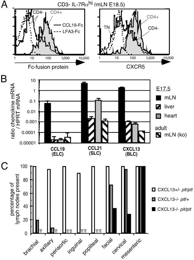
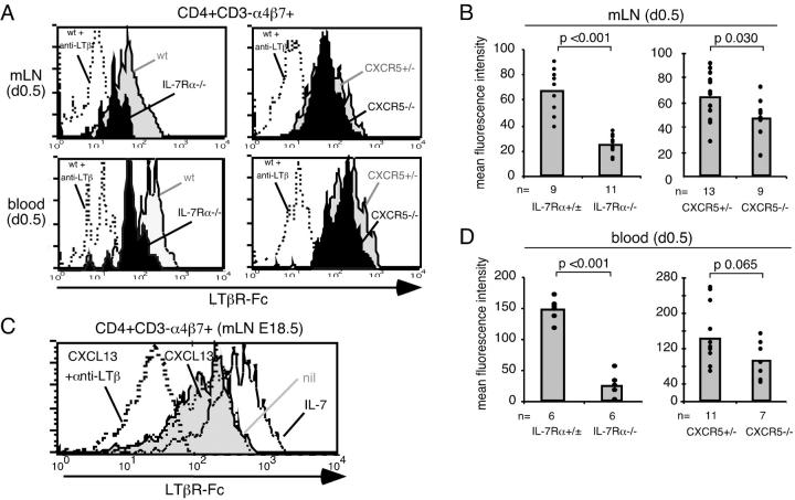
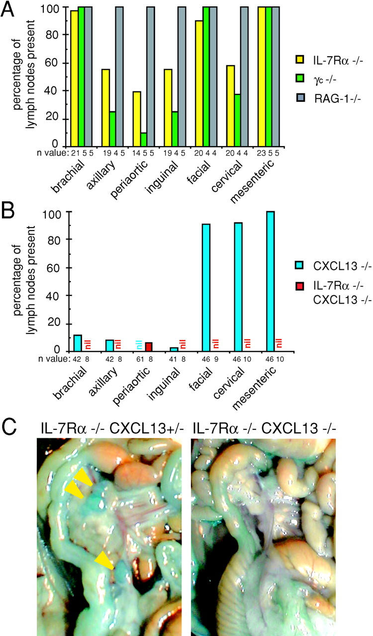
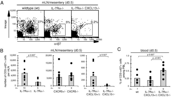
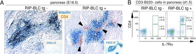
Lymphoid tissue development is associated with local accumulation of CD4+ CD3- IL-7R alpha hi hematopoietic cells that deliver lymphotoxin (LT)alpha 1 beta 2 signals to resident stromal cells. Previous studies have established an important role for CXCL13 (BLC) in the development of Peyer's patches (PP) and some peripheral lymph nodes (LNs), but the chemokine requirements for several LN types, including mesenteric LNs, remain undefined. Using CXCL13-/- mice that additionally carry the paucity of LN T cell mutation (plt/plt), we discovered that CCR7 ligands function in peripheral LN development. We also tested for a genetic interaction during LN development between CXCL13 and a cytokine receptor required in PP development, IL-7R alpha. Mice deficient for both CXCL13 and IL-7R alpha displayed a striking absence of LNs, including mesenteric LNs. These data extend the role of CXCL13 to the development of all LNs and establish a previously unappreciated role for IL-7R alpha in this process. Both circulating and LN CD4+ CD3- IL-7R alpha hi cells are shown to express LT alpha 1 beta 2 in an IL-7R alpha-dependent manner. Furthermore, CXCL13 was found to be sufficient to mediate CD4+ CD3- IL-7R alpha hi cell recruitment in vivo to an ectopic site. These findings indicate that CXCL13 and CCR7 ligands promote accumulation of CD4+ CD3- IL-7R alpha hi cells, delivering IL-7R alpha-dependent LT alpha 1 beta 2 signals critical for LN development.


Figures

Figure 1.
Figure 1.
The chemokines CXCL13 (BLC), CCL19 (ELC), and CCL21 (SLC) play overlapping roles in the development of facial, cervical, brachial, and axillary LNs. (A) Representative histograms showing the staining with CCL19-Fc fusion protein (left) or CXCR5 on the CD4+ (shaded) and CD4 (open) subset of CD3 IL-7Rαhi cells from E 18.5 mesenteric LNs (mLN) of wild-type mice. As a negative control, staining with the fusion protein hLFA3-Fc is shown for embryonic CD4 CD3 IL-7Rαhi cells (left) and CXCR5 staining is shown for CD4 CD3 IL-7Rα triple negative cells (TN, right) in dotted lines. CD3 indicates low or negative for the lineage markers B220, CD3, CD11c, and CD11b. (B) Quantitative PCR analysis of chemokine expression in the indicated organs from E17.5 B6 mice. As negative controls, mesenteric LN samples from adult CCL19/CCL21-ser (plt/plt) or CXCL13-deficient mice were used. CCL21 expression in fetal heart and the weak expression in fetal liver and in plt/plt mesenteric LNs is likely due to lymphatic expression of the second CCL21 gene, CCL21-leu. Three samples were analyzed per group except for the CXCL13−/− mesenteric LN. ND, not determined. (C) Quantitative assessment of the presence of the indicated LN types detected macroscopically after injection of Chicago sky blue dye (refer to Materials and Methods) in littermate mice deficient for CCL19/CCL21-ser (plt/plt), CXCL13, or CCL19/CCL21-ser and CXCL13 combined. 10–13 mice were investigated per group.
Figure 2.
Figure 2.
IL-7Rα but not CXCR5 signals promote induction of LTα1β2 on CD4+ CD3 α4β7+ cells in developing mesenteric LNs and in blood. (A) Representative histograms showing LTβR-Fc staining on CD4+ CD3 α4β7+ cells from day 0.5 mesenteric LNs (mLN) or blood of wild-type (wt) or CXCR5+/− (shaded) and IL-7Rα−/− or CXCR5−/− mice (solid) as indicated. As a negative control, LTβR-Fc staining of wild-type cells pretreated with anti-LTβ blocking antibody is shown (dotted line). (B) Compilation of data as shown in A, showing the mean fluorescence intensity (MFI) of LTβR-Fc staining on CD4+ CD3 α4β7+ cells from mesenteric LNs of newborn mice of the indicated genotypes. (C) LTβR-Fc staining of sorted CD4+ CD3 α4β7+ cells from mesenteric LN cells of E18.5 mice that had been cultured for 6 h in vitro without treatment (nil, shaded), with 2 μg/ml CXCL13 (thin dotted line), or with 20 ng/ml IL-7 (open). As a negative control, LTβR-Fc staining of wild-type cells treated with anti-LTβ blocking antibody is shown (thick dotted line). CD3 indicates low or negative for the lineage markers B220, CD3, CD11c, and CD11b. Data are representative of two experiments. Similar observations were made for CD4 CD3 α4β7+ cells in the same cultures. (D) Compilation of data as in A, showing the MFI of LTβR-Fc staining on CD4+ CD3 α4β7+ cells from blood of newborn mice of the indicated genotypes. In B and D, plotted values represent the MFI after subtraction of the background LTβR-Fc staining on cells treated with LTβ blocking antibody. The data are compiled from two to four independent experiments, with littermate mice used as controls. IL-7Rα−/− mice were either CXCL13+/+ or CXCL13+/−, and data were pooled as no difference was observed between these two groups.
Figure 3.
Figure 3.
Deficiency of macroscopically detectable LNs in IL-7Rα−/− and IL-7Rα−/− CXCL13−/− mice. (A and B) Quantitative assessment of the presence of the indicated LNs in mice deficient for IL-7Rα, γc chain, RAG-1 (A), CXCL13, or IL-7Rα and CXCL13 combined (B), compared with wild-type levels (100%). Importantly, with the exception of one periaortic LN, all LNs were missing in IL-7Rα−/− CXCL13−/− mice. The LN enumeration data for CXCL13−/− mice is equivalent to that previously reported (reference 11) and is shown here to facilitate the comparison with the other mouse strains. No difference in LN presence was observed between IL-7Rα−/− CXCL13+/+ and IL-7Rα−/− CXCL13+/− mice. 100% of IL-7Rα−/− mice also lacked sacral LNs (not depicted). The n value indicates the number of mice that were investigated using Chicago sky blue dye. (C) Representative photograph of mesentery region in IL-7Rα−/− CXCL13+/− and IL-7Rα−/− CXCL13−/− mice that had been injected with Chicago sky blue dye, showing the presence of several small mesenteric LNs in IL-7Rα−/− CXCL13+/− mice (arrow heads) and the absence of detectable mesenteric LNs in IL-7Rα−/− CXCL13−/− mice.
Figure 4.
Figure 4.
IL-7Rα and CXCR5 signals promote the accumulation of CD3 α4β7+ cells in developing mesenteric LNs. (A) Representative dot plots of day 0.5 mesenteric LNs or mesentery region from wild-type, IL-7Rα−/−, and IL-7Rα−/− CXCL13−/− mice showing reduced numbers and absence of CD3 α4β7+ cells in IL-7Rα−/− and IL-7Rα−/− CXCL13−/− mice, respectively. (B) Histograms depicting the number of CD3 α4β7+ cells per mesenteric LN of newborn mice of the genotype indicated. In each case, 40% of CD3 α4β7+ cells were CD4+. (C) Histogram showing the percentage of CD3 α4β7+ cells in peripheral blood leukocytes of newborn mice of the genotype indicated. n, number of mice investigated. Each set is a compilation of two to three different experiments, with littermate mice used as controls. In B and C, CD3 indicates low or negative for the lineage markers B220, CD3, CD11c, and CD11b except for IL-7Rα+/− mice (B) where only B220, CD3, and CD11c were used as lineage markers.
Figure 5.
Figure 5.
CD4+ IL-7Rαhi cells in the pancreas of RIP-CXCL13 transgenic mice. (A) Immunohistochemical analysis of pancreas from RIP-CXCL13 transgenic (tg+) and littermate control (tg−) E 18.5 embryos to detect insulin-producing β cells (blue) and CD4+ cells (brown). Insets, adjacent sections stained to detect CXCL13 transgene expression (blue). (B) Frequency of IL-7Rαhi CD3 B220 cells in day 1.5 pancreas tissue of transgenic and littermate control mice. Numbers show average percentages of CD4+ and CD4 IL-7Rαhi cells ± standard deviation in seven tg− and five tg+ mice.

References

    1. Fu, Y.-X., and D.D. Chaplin. 1999. Development and maturation of secondary lymphoid tissues. Annu. Rev. Immunol. 17:399–433. - PubMed
    1. Mebius, R.E., P. Rennert, and I.L. Weissman. 1997. Developing lymph nodes collect CD4+CD3− LTbeta+ cells that can differentiate to APC, NK cells, and follicular cells but not T or B cells. Immunity. 7:493–504. - PubMed
    1. Nishikawa, S., K. Honda, H. Hashi, and H. Yoshida. 1998. Peyer's patch organogenesis as a programmed inflammation: a hypothetical model. Cytokine Growth Factor Rev. 9:213–220. - PubMed
    1. Fukuyama, S., T. Hiroi, Y. Yokota, P.D. Rennert, M. Yanagita, N. Kinoshita, S. Terawaki, T. Shikina, M. Yamamoto, Y. Kurono, et al. 2002. Initiation of NALT organogenesis is independent of the IL-7R, LTβR, and NIK signaling pathways but requires the Id2 gene and CD3− CD4+CD45+ cells. Immunity. 17:31–40. - PubMed
    1. Finke, D., H. Acha-Orbea, A. Mattis, M. Lipp, and J. Kraehenbuhl. 2002. CD4(+)CD3(−) cells induce Peyer's patch development. Role of alpha4beta1 integrin activation by CXCR5. Immunity. 17:363–373. - PubMed

Publication types