. 2003 May 27;100(11):6813-8.
doi: 10.1073/pnas.1137276100. Epub 2003 May 5.

Alcohol potently inhibits the kainate receptor-dependent excitatory drive of hippocampal interneurons

Affiliations

Alcohol potently inhibits the kainate receptor-dependent excitatory drive of hippocampal interneurons

Mario Carta et al. Proc Natl Acad Sci U S A. .

Abstract

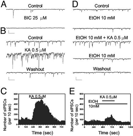
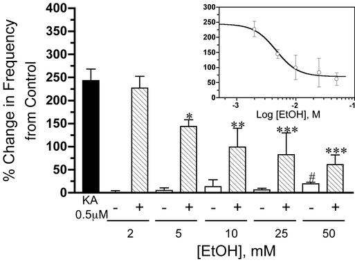
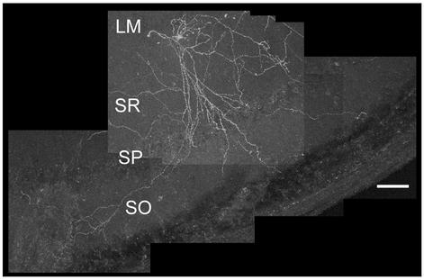
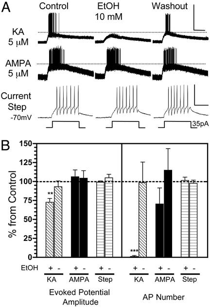
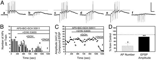
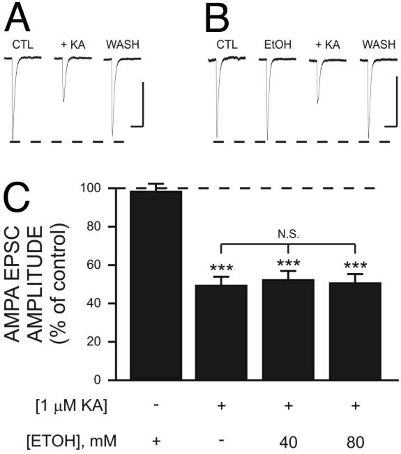
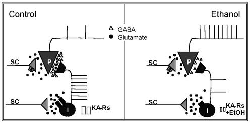
Kainate receptors (KA-Rs) are members of the glutamate-gated family of ionotropic receptors, which also includes N-methyl-d-aspartate (NMDA) and alpha-amino-3-hydroxy-5-methylisoxazole-4-propionate (AMPA) receptors. KA-Rs are important modulators of interneuron excitability in the CA1 region of the hippocampus. Activation of these receptors enhances interneuron firing, which robustly increases spontaneous inhibitory postsynaptic currents in pyramidal neurons. We report here that ethanol (EtOH) potently inhibits this KA-R-mediated effect at concentrations as low as those that can be achieved in blood after the ingestion of just 1-2 drinks (5-10 mM). Pressure application of kainate, in the presence of AMPA and NMDA receptor antagonists, evoked depolarizing responses in interneurons that triggered repetitive action potential firing. EtOH potently inhibited these responses to a degree that was sufficient to abolish action potential firing. This effect appears to be specific for KA-Rs, as EtOH did not affect action potential firing triggered by AMPA receptor-mediated depolarizing responses. Importantly, EtOH inhibited interneuron action potential firing in response to KA-R activation by synaptically released glutamate, suggesting that our findings are physiologically relevant. KA-R-dependent modulation of glutamate release onto pyramidal neurons was not affected by EtOH. Thus, EtOH increases excitability of pyramidal neurons indirectly by inhibiting the KA-R-dependent drive of gamma-aminobutyric acid (GABA)ergic interneurons. We postulate that this effect may explain, in part, some of the paradoxical excitatory actions of this widely abused substance. The excitatory actions of EtOH may be perceived as positive by some individuals, which could contribute to the development of alcoholism.


Figures

Fig. 1.
Fig. 1.
EtOH inhibits the KA-R-mediated increase of sIPSC frequency in CA1 pyramidal neurons. (A) sIPSCs are fully blocked by the GABAA-R antagonist, bicuculline (BIC; 25 μM). (B and C) Bath application of KA (0.5 μM) for 3.5 min, in the presence of GYKI 53665 (30 μM) and DL-AP5 (100 μM), induced a robust and reversible sIPSC frequency increase. (D and E) EtOH (10 mM) did not affect either the basal amplitude or frequency of sIPSCs, but reduced the increase in frequency induced by KA-R-activation. (Scale bars: 100 pA and 250 ms.)
Fig. 2.
Fig. 2.
EtOH inhibits the KA-R-mediated effect on sIPSC frequency in a concentration-dependent manner. The filled bar represents the average percent change induced by KA (0.5 μM) on sIPSC frequency. Striped bars represent the effect of increasing concentrations of EtOH on this KA-R-dependent effect. Open bars represent the percent change in basal sIPSC frequency induced by EtOH alone. (Inset) A nonlinear regression fit of the inhibitory effect of EtOH on the KA-R-induced increase of sIPSC frequency (IC50 = 4.6 mM). Each bar or symbol represents the mean ± SEM of 7–28 neurons. *, P < 0.05; **, P < 0.01; ***, P < 0.001, by one-way ANOVA followed by Bonferroni's multiple comparison test; #, P < 0.05, by one sample t test versus theoretical mean of zero.
Fig. 3.
Fig. 3.
Anatomical reconstruction of a stratum radiatum-stratum lacunosum moleculare interneuron. Shown is a single z axis projection of 20 confocal microscopy sections (4 μm) of one of the interneurons that were studied electrophysiologically. See Materials and Methods for details of histochemical procedures. Similar results were obtained with six additional neurons (data not shown). (Scale bar: 100 μm.) L-M, stratum lacunosum moleculare; SR, stratum radiatum; SP, stratum pyramidale; SO, stratum oriens.
Fig. 4.
Fig. 4.
EtOH inhibits evoked potentials and AP firing triggered by pressure application of KA onto interneurons. (A) Sample traces of current–clamp recordings (I = 0) from CA1 stratum radiatum-stratum lacunosum moleculare interneurons. (Top) In the presence of DL-AP5 (100 μM) and GYKI 53655 (30 μM), pressure application of KA (5 μM in micropipette located ≈200 μm from the soma) induced reproducible evoked potentials and bursts of AP firing. Bath application of EtOH (10 mM) induced a reversible decrease of the KA-R-mediated evoked potentials and also abolished firing. (Middle) The same experiment was performed by pressure-delivering 5 μM AMPA to another interneuron in the presence of DL-AP5 only. EtOH did not inhibit AMPAR-mediated evoked potential amplitude and AP firing. (Bottom) Lack of an effect of EtOH on depolarization-induced AP firing in another interneuron. (Scale bars for Top and Middle: 50 mV and 5 sec; for Bottom: 50 mV and 100 msec.) (B) Summary of the effects of EtOH on KA-R-dependent and AMPAR-dependent evoked potential amplitude and AP number. Also shown is the summary of the effect of EtOH on the amplitude of depolarization induced by current injection and AP number in response to this current injection. Data were normalized with respect to control responses (represented by the dashed line). +, the effect of 10 mM EtOH; -, the effect of the washout. Each bar represents the mean ± SEM of five to eight neurons. **, P < 0.01, ***, P < 0.001, by one sample t test versus theoretical mean of 100.
Fig. 5.
Fig. 5.
EtOH inhibits KA-R-mediated interneuron EPSPs and AP firing evoked by stimulation of the Schaffer collaterals. (A) Sample traces of current–clamp recordings (I = 0) from a CA1 stratum radiatum-stratum lacunosum moleculare interneuron. (a) In the presence of DL-AP5 (100 μM), bicuculline (25 μM), and SCH-50911 (20 μM), a train of five stimuli (20 Hz) delivered in the stratum radiatum induced non-NMDA EPSPs that triggered a burst of APs. (b) GYKI 53665 (30 μM) blocked the AMPA component of the EPSPs and eliminated firing. (c) An elevation in the stimulation intensity increased the amplitude of the KA-R-mediated EPSPs and restored AP firing. (d and e) Bath application of EtOH (10 mM) reversibly decreased the peak amplitude of the KA-R-mediated compound EPSPs and reduced AP number. (f) The KA-R-mediated EPSPs were fully blocked by DNQX (80 μM). (Scale bars: 50 mV and 100 msec.) (B and C) Time courses illustrating the effects of the sequential application of GYKI, EtOH, and DNQX on EPSP amplitude and AP number in the same cell illustrated in A. (D) Summary of the effect of EtOH (10 mM) on AP number and EPSP amplitude. Each bar represents the mean ± SEM of five neurons. *, P < 0.05; **, P < 0.01, by one sample t test versus theoretical mean of 100. Data were normalized with respect to control responses (represented by the dashed line).
Fig. 6.
Fig. 6.
EtOH has no effect on presynaptic KA receptor-dependent inhibition of AMPA EPSCs in CA1 pyramidal neurons. Traces are averages of five to nine AMPA EPSCs recorded in the presence of bicuculline (20 μM) and D-AP5 (50 μM). Representative traces illustrating the inhibitory effect of 1 μM KA on AMPAR-mediated EPSCs in the absence (A) and presence (B) of 80 mM EtOH. Note that EtOH alone has no effect on the amplitude of AMPA EPSCs or KA inhibition of AMPA EPSCs. (Scale bars: 100 pA and 200 msec.) (C) Summary of the effect of EtOH on KA inhibition of AMPA EPSCs. ***, P < 0.001 with respect to responses obtained in the presence of EtOH alone by one-way ANOVA; each bar represents the mean ± SEM of 7–27 neurons. Data were normalized with respect to control responses (represented by the dashed line).
Fig. 7.
Fig. 7.
Simplified model of the EtOH-induced inhibition of the KA-R-mediated excitatory drive of interneurons. (Left) Under control conditions, glutamate (circles) released from the Schaffer collaterals (SC) activates interneuronal KA-Rs, which depolarize interneurons (I), and induces AP firing. This effect triggers sIPSCs onto CA1 pyramidal neurons (P); GABA is represented by triangles. Presynaptic KA-Rs in Schaffer collateral glutamatergic terminals that synapse onto CA1 pyramidal neurons are depicted in gray. Activation of these receptors inhibits glutamate release. The net effect of KA-Rs activation is to increase tonic inhibition of CA1 pyramidal neuron's excitability. AMPARs and axonal KA-Rs are not depicted for clarity. (Right) In the presence of low concentrations of EtOH (5–10 mM), KA-Rs in interneurons are inhibited, which reduces the excitatory drive to these neurons, reduces GABAA-R-mediated sIPSCs, and increases the excitability of pyramidal neurons. KA-Rs in glutamatergic terminals are unaffected by EtOH. The effects of higher concentrations of EtOH (≥20 mM) are not depicted for clarity (see Discussion); at these concentrations, EtOH will (i) reduce the inhibitory effect of KA-R activation on eIPSCs (15), (ii) inhibit KA-R-mediated synaptic currents in CA3 pyramidal neurons (24), and (iii) modulate the function of other neuronal proteins, including GABAA-Rs and NMDARs.

References

    1. Kullmann, D. M. (2001) Neuron 32, 561-564. - PubMed
    1. Schmitz, D., Mellor, J., Frerking, M. & Nicoll, R. A. (2001) Proc. Natl. Acad. Sci. USA 98, 11003-11008. - PMC - PubMed
    1. Castillo, P. E., Malenka, R. C. & Nicoll, R. A. (1997) Nature 388, 182-186. - PubMed
    1. Bureau, I., Bischoff, S., Heinemann, S. F. & Mulle, C. (1999) J. Neurosci. 19, 653-663. - PMC - PubMed
    1. Kamiya, H. & Ozawa, S. (1998) J. Physiol. (London) 509, 833-845. - PMC - PubMed

Publication types

Substances

LinkOut - more resources