. 2003 May 27;100(11):6628-33.
doi: 10.1073/pnas.1131932100. Epub 2003 May 5.

Subcellular localization of yeast ribonucleotide reductase regulated by the DNA replication and damage checkpoint pathways

Affiliations

Subcellular localization of yeast ribonucleotide reductase regulated by the DNA replication and damage checkpoint pathways

Ruojin Yao et al. Proc Natl Acad Sci U S A. .

Abstract

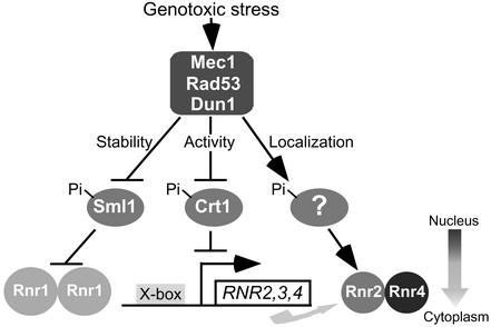
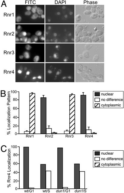
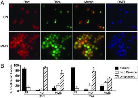
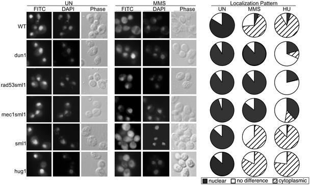
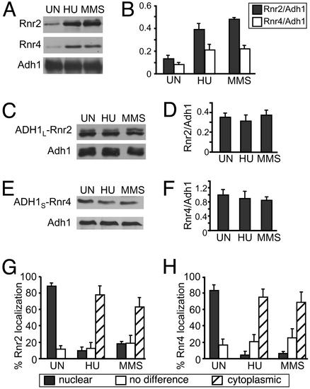
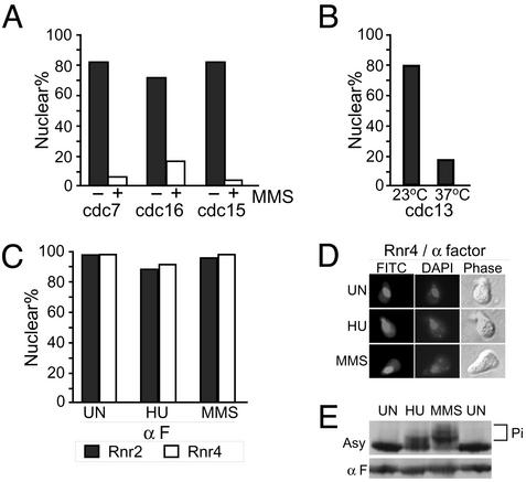
The fidelity of DNA replication and repair processes is critical for maintenance of genomic stability. Ribonucleotide reductase (RNR) catalyzes the rate-limiting step in dNTP production and thus plays an essential role in DNA synthesis. The level and activity of RNR are highly regulated by the cell cycle and DNA damage checkpoints, which maintain optimal dNTP pools required for genetic fidelity. RNRs are composed of a large subunit that binds the nucleoside diphosphate substrates and allosteric effectors and a small subunit that houses the di-iron tyrosyl radical cofactor essential for the reduction process. In Saccharomyces cerevisiae, there are two large subunits (Rnr1 and Rnr3) and two small subunits (Rnr2 and Rnr4). Here we report the subcellular localization of Rnr1-4 during normal cell growth and the redistribution of Rnr2 and Rnr4 in response to DNA damage and replicational stress. During the normal cell cycle, Rnr1 and Rnr3 are predominantly localized to the cytoplasm and Rnr2 and Rnr4 are predominantly present in the nucleus. Under genotoxic stress, Rnr2 and Rnr4 become redistributed to the cytoplasm in a checkpoint-dependent manner. Subcellular redistribution of Rnr2 and Rnr4 can occur in the absence of the transcriptional induction of the RNR genes after DNA damage and likely represents a posttranslational event. These results suggest a mechanism by which DNA damage checkpoint modulates RNR activity through the temporal and spatial regulation of its subunits.


Figures

Fig. 1.
Fig. 1.
A schematic representation of different mechanisms involved in the regulation of RNR activity by the DNA damage checkpoint pathways.
Fig. 2.
Fig. 2.
The large and small subunits of RNR are localized in distinctive subcellular compartments. (A) Subcellular localization of RNR subunits. For Rnr3 staining, crt1 mutant cells were used. (B) Quantification of RNR subcellular localization. Three independent cultures were processed for IMF analysis. For each experiment, 200 cells were counted. Percentages of cells with distinct localization patterns were represented as follows: dark bar, cells with a predominantly nuclear signal; hatched bar, cells with a predominantly cytoplasmic signal; open bar, cells with ubiquitous fluorescence in both the nucleus and the cytoplasm (no difference); error bars, SD. (C) Quantification of Rnr4 localization in cells synchronized in S phase. WT and dun1 cells were released from α-factor-mediated G1 block and harvested in mid-S phase ( min after release) for IMF. Similar results were observed for Rnr2. The symbols for bar representation are the same as in B.
Fig. 3.
Fig. 3.
RNR small subunits become redistributed under genotoxic stress. (A) Colocalization of Rnr1 and Rnr4 in the cytoplasm after MMS treatment. An HA-RNR1 strain was grown to early log phase and split in half, one half was left untreated (UN) and the other was treated with 0.02% MMS for 2 h. Cells were processed for IMF by using a mouse anti-HA and a rabbit anti-Rnr4. Cy3-conjugated (red) goat-anti-mouse and FITC-conjugated (green) goat-anti-rabbit Abs were used to detect HA-Rnr1 and Rnr4, respectively. DNA was visualized by DAPI (blue). (B) Quantification of Rnr2 and Rnr4 localization changes after HU and MMS treatment. Counting of IMF and the symbols for bar representation were the same as in the Fig. 2 legend.
Fig. 4.
Fig. 4.
RNR small subunit redistribution is regulated by the DNA damage checkpoint kinases. Early log-phase cultures were treated with 150 mM HU or 0.02% MMS for 2 h and then processed for IMF. Shown here are the results of Rnr4 (similar results were also observed for Rnr2). Isogenic strains were dun1::HIS3, rad53::HIS3 sml1::KAN, mec1::HIS3 sml1::KAN, sml1::KAN, and hug1::HIS3. Each experiment was performed in duplicate with 200 cells counted. Each pie chart represents the percentage of cells with a specific IMF pattern: dark, nuclear; hatched, cytoplasmic; white, no difference between the nucleus and the cytoplasm.
Fig. 5.
Fig. 5.
DNA damage-induced RNR small subunit redistribution is independent of transcriptional induction. (A and B) Induction of endogenous Rnr2 and Rnr4 by HU and MMS. Early log-phase cultures were treated with 150 mM HU or 0.02% MMS for 2 h and processed for Western blot by using anti-Rnr2 and anti-Rnr4. Two isoforms of Rnr2 were observed. The same blot was reprobed with anti-Adh1 to visualize Adh1, which served as a loading control. Analysis was performed by densitometry, correcting Rnr2 and Rnr4 levels for Adh1. Each bar (black, Rnr2; white, Rnr4) is the mean of the samples shown in A plus two additional blots (n = 3). (C and D) No induction of ADH1 promoter-driven Rnr2 by either HU or MMS. A 3MYC-RNR2 driven by a 1.1-kb ADH1 promoter (ADH1L-RNR2) was introduced into an rnr2 deletion strain. Cells were treated with HU and MMS, and Rnr2 levels were detected by anti-Myc and normalized to Adh1 signals on the same blot. (E and F) No induction of ADH1 promoter-driven Rnr4 by either HU or MMS. An HA-RNR4 driven by a 0.4-kb ADH1 promoter (ADH1S-RNR4) was introduced into an rnr4 deletion strain. Cells were treated with HU and MMS, and Rnr4 levels were detected by anti-HA and normalized to Adh1 signal on the same blot. (G and H) Quantification of ADH1L-Rnr2 (G) and ADH1S-Rnr4 (H) localization after genotoxic stress. The symbols for bar representation are the same as in the Fig. 2 legend.
Fig. 6.
Fig. 6.
Cell cycle effect on Rnr2 and Rnr4 redistribution induced by genotoxic stress. (A) Change of Rnr4 nuclear localization in cdc mutants treated with MMS. cdc7-1, cdc16-123, and cdc15-2 cells were arrested at 37°C for 2 h, treated with 0.02% MMS for an additional 2 h, and processed for IMF. The percentage of cells displaying a predominantly nuclear Rnr4 signal was calculated by counting 200 cells per experiment, each done in duplicate. (B) Loss of Rnr4 nuclear localization induced by cdc13-1-mediated DNA damage. A cdc13-1 culture was grown at 23°C to early log phase and shifted to 37°C for 2 h. Cells were quantified as described in A. (C) No change in Rnr2 and Rnr4 localization in G1 cells treated with HU or MMS. Asynchronous cultures were blocked in G1 with α-factor (α F). The arrest was maintained while cells were treated with 150 mM HU or 0.02% MMS for 2 h, and then processed for IMF. The percentages of each cell population with a predominantly nuclear signal for Rnr2 (black bar) or Rnr4 (white bar) were quantified as described in A.(D) A representative collage of Rnr4 IMF images in MMS-treated, α-factor-arrested G1 cells. (E) Comparison of HU- and MMS-induced Rad53 phosphorylation between cells in an asynchronous (Asy) culture and G1-arrested (α F) cells.

References

    1. Zhou, B. B. & Elledge, S. J. (2000) Nature 408, 433–439. - PubMed
    1. Khanna, K. K. & Jackson, S. P. (2001) Nat. Genet. 27, 247–254. - PubMed
    1. Kolodner, R. D., Putnam, C. D. & Myung, K. (2002) Science 297, 552–577. - PubMed
    1. Lengauer, C., Kinzler, K. W. & Vogelstein, B. (1998) Nature 396, 643–649. - PubMed
    1. Abraham, R. T. (2001) Genes Dev. 15, 2177–2196. - PubMed

Publication types

MeSH terms

Substances