. 2003 May 13;100(10):5840-5.
doi: 10.1073/pnas.1036475100. Epub 2003 May 5.

Mesenchymal progenitor self-renewal deficiency leads to age-dependent osteoporosis in Sca-1/Ly-6A null mice

Affiliations

Mesenchymal progenitor self-renewal deficiency leads to age-dependent osteoporosis in Sca-1/Ly-6A null mice

Mortaza Bonyadi et al. Proc Natl Acad Sci U S A. .

Abstract

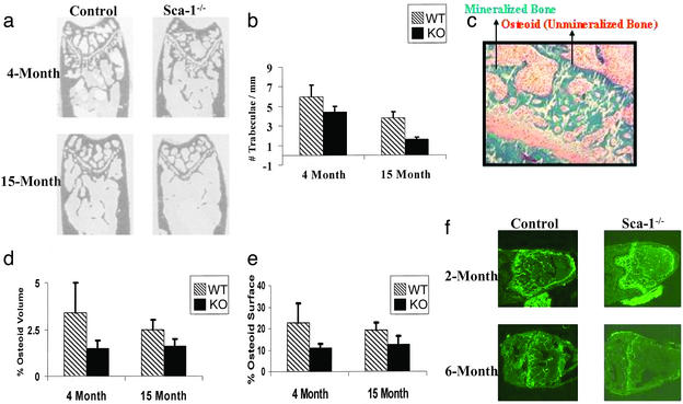
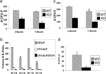
The cellular and molecular mechanisms that underlie age-dependent osteoporosis, the most common disease in the Western Hemisphere, are poorly understood in part due to the lack of appropriate animal models in which to study disease progression. Here, we present a model that shows many similarities to the human disease. Sca-1, well known for its expression on hematopoietic stem cells, is present on a subset of bone marrow stromal cells, which potentially include mesenchymal stem cells. Longitudinal studies showed that Sca-1(-/-) mice undergo normal bone development but with age exhibit dramatically decreased bone mass resulting in brittle bones. In vivo and in vitro analyses demonstrated that Sca-1 is required directly for the self-renewal of mesenchymal progenitors and indirectly for the regulation of osteoclast differentiation. Thus, defective mesenchymal stem or progenitor cell self-renewal may represent a previously uncharacterized mechanism of age-dependent osteoporosis in humans.


Figures

Figure 1
Figure 1
BMD and mechanical analysis of Sca-1−/− (KO) and Sca-1+/+ mice (WT) demonstrate that Sca-1−/− mice exhibit age-related osteoporosis. (a) Significantly higher (P < 0.0001) whole-body BMD was observed in control mice at 12 months of age compared with Sca-1−/− animals, whereas no significant differences were observed at 2 months of age. (b) The BMD of excised femurs and L6 vertebrae at 15 months shows significantly less mineralization in Sca-1−/− femurs and vertebrae than those of WT mice, with a greater difference in the vertebrae. (c) Vertebral stress-strain response at 15 months demonstrated that vertebrae from KO were substantially weaker and more compliant than bones from WT mice. (d) Femoral stress–strain response at 15 months showed that femurs from KO were more brittle and weak than femurs from controls, although equally as stiff.
Figure 2
Figure 2
Sca-1−/− mice have a non-cell autonomous osteoclast defect. (a) The absolute number of TRAP+ (osteoclasts) cells in femurs of both 2- and 9-month-old Sca-1−/− mice is <40% of that in Sca-1+/+ mice. (b) Osteoblast (OB)/splenocytes (monocyte, MO) coculture experiments. Sca-1+/+ (WT) osteoblast (2 × 10⁴ cells per well) and splenic (1.5 × 10⁵ cells per well) cultures were used to standardize the culture results (i.e., 100% on x axis); osteoclast formation in all other culture combinations is specified as the percentage compared with the WT cocultures. The number of TRAP+ cells in cocultures in which monocytes are derived from WT mice and osteoblasts are derived from either Sca-1−/− or WT mice is significantly different (P < 0.001). However, osteoclast formation is not significantly altered in cocultures in which OB are derived from KO and monocytes from either Sca-1−/− or WT mice (P = 0.4). (c) The number of mononucleated, binucleated, and multinucleated osteoclasts, and total number of osteoclasts (TRAP+ cells) generated in vitro by different combinations of osteoblasts and monocytes derived from WT or Sca-1−/− mice. (d) Images of TRAP+ mononucleated, binucleated, and multinucleated cells generated by a combination of monocytes and osteoblasts derived from Sca-1−/− mice in culture.
Figure 3
Figure 3
Progressive diminution of bone mass in Sca-1−/− mice. (a) Plastic sections of femurs from Sca-1−/− and Sca-1+/+ mice at 4 and 15 months of age used for image analysis. (b) Image analysis demonstrates that Sca-1−/− femurs have 27% (P < 0.001) fewer trabeculae compared with control bones at 4 months of age and 58% (P < 0.001) compared with Sca-1+/+ bones at 15 months of age. (c) Goldner's trichrome staining of unmineralized bone sections of femur showing mineralized bone (green), osteoid (red), and cartilage (pink) was used to measure osteoid volume and osteoid surface. (d and e) The percent osteoid volume and osteoid surface calculated from unmineralized femoral sections from 4- and 15-month-old Sca-1−/− mice are less than those of Sca-1+/+ mice. (f) Double calcein labeling of bones from Sca-1−/− and Sca-1+/+ mice at 2 and 6 months of age revealed that bone formation in Sca-1−/− mice is essentially normal at 2 months of age but is dramatically reduced by 6 months of age.
Figure 4
Figure 4
In vitro analysis of the cellular and molecular basis of the bone formation deficiency in Sca-1−/− mice. (a) The numbers of CFU-F and CFU-ALP generated in BM cultures derived from 2-month-old Sca-1−/− mice are <50% of those derived from Sca-1+/+ mice. (b) The number of CFU-O formed from Sca-1−/− cultures is ≈50% of CFU-O from Sca-1+/+ cultures. (c) The number of ALP+ cells in calvarial cultures from Sca-1−/− at different time points is consistently less than that of Sca-1+/+ mice. The cultures were initiated with 10,000 cells. (d) RT-PCR analysis of osteoblast- and adipocyte-associated genes during in vitro differentiation at 3, 6, and 10 days excludes the possible disturbances of osteoblast and/or adipocyte pathways in Sca-1−/− mice. Expression of adipsin (mADP), alkaline phosphatase (ALP), osteopontin (OPN), Sca-1, and bone sialoprotein (BSP) genes were examined, and L32 expression was used as a control for relative mRNA levels.
Figure 5
Figure 5
Mesenchymal progenitor self-renewal deficiency in Sca-1−/− mice. (a) Total number of CFU-F formed in Sca-1−/− compared with Sca-1+/+ cultures is reduced 45% and 67%, respectively, in 2- and 7-month-old mice. (b) The total numbers of CFU-F and CFU-ALP are decreased in primary BM stromal cultures derived from 2- and 7-month-old Sca-1−/− cultures; however, the ratio of CFU-ALP/CFU-F is consistent (≈10%) between KO and WT cultures, indicating that the decrease in osteoprogenitors is due to a reduced total number of stroma progenitors in Sca-1−/− mice. (c) Total number of CFU-F in secondary BM stromal cultures derived from 2- and 7-month-old Sca-1−/− mice is ≈50% of those derived from Sca-1+/+ mice, suggesting a self-renewal deficiency by Sca-1−/− stromal progenitors. (d) Reduced CFU-A (adipocyte) was generated in BM stromal cultures derived from Sca-1−/− mice.

References

    1. Wasnich R D. In: Primer on the Metabolic Bone Diseases and Disorders of Mineral Metabolism. Favus M J, editor. Philadelphia: Lippincott Williams & Wilkins; 1996. pp. 249–251.
    1. Bellows C G, Wang Y H, Heersche J N M, Aubin J E. Endocrinology. 1994;134:2221–2229. - PubMed
    1. Aubin J E. J Cell Biochem. 1998;Suppl. 30/31:73–82. - PubMed
    1. Roodman G D. Exp Hematol. 1999;27:1229–1241. - PubMed
    1. Spangrude G J, Heimfeld S, Weissman I L. Science. 1988;241:58–62. - PubMed

Publication types

MeSH terms