. 2003 May 13;100(10):5646-51.
doi: 10.1073/pnas.0836054100. Epub 2003 May 5.

Binding and recognition in the assembly of an active BRCA1/BARD1 ubiquitin-ligase complex

Affiliations

Binding and recognition in the assembly of an active BRCA1/BARD1 ubiquitin-ligase complex

Peter S Brzovic et al. Proc Natl Acad Sci U S A. .

Abstract

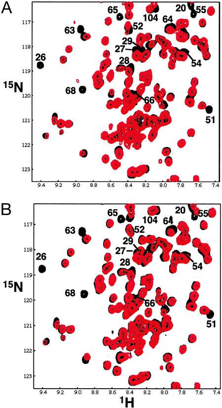
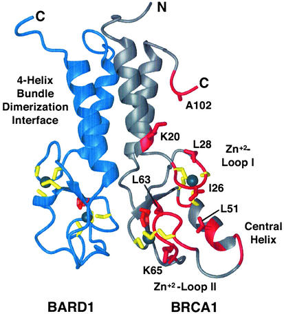
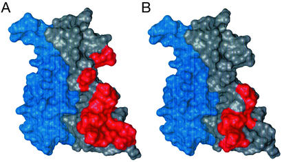
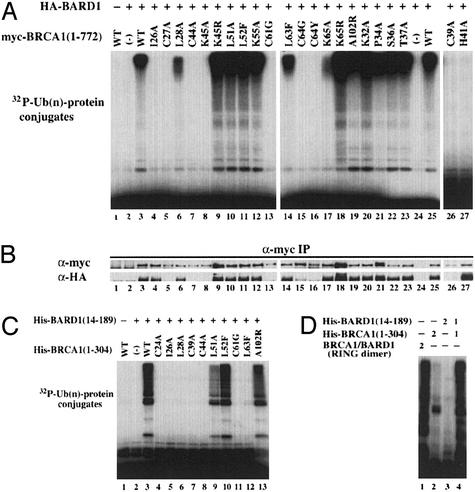
BRCA1 is a breast and ovarian cancer tumor suppressor protein that associates with BARD1 to form a RINGRING heterodimer. The BRCA1BARD1 RING complex functions as an ubiquitin (Ub) ligase with activity substantially greater than individual BRCA1 or BARD1 subunits. By using NMR spectroscopy and site-directed mutagenesis, we have mapped the binding site on the BRCA1BARD1 heterodimer for the Ub-conjugating enzyme UbcH5c. The results demonstrate that UbcH5c binds only to the BRCA1 RING domain and not the BARD1 RING. The binding interface is formed by the first and second Zn(2+)-loops and central alpha-helix of the BRCA1 RING domain, a region disrupted by cancer-predisposing mutations. Unexpectedly, a second Ub-conjugating enzyme, UbcH7, also interacts with the BRCA1BARD1 complex with similar affinity, although it is not active in Ub-ligase activity assays. Thus, binding alone is not sufficient for BRCA1-dependent Ub-ligase activity.


Figures

Figure 1
Figure 1
NMR titrations of E2 binding to the BRCA1/BARD1 heterodimer. Overlay of an expanded region of 2D 1H–15N TROSY spectra of 15N-labeled BRCA1/BARD1 heterodimer with 0 (black spectra) and 0.5 (red spectra) equivalent unlabeled UbcH5c (A) or UbcH7 (B). Resonances significantly perturbed on the addition of UbcH5c (K20, I26, C27, L28, L51, L52, Q54, K55, L63, C64, K65, N66, I68, and S104 of BRCA1) are labeled. Other perturbed resonances (C24, K45, F46, C61, and A102) lie outside this spectral region.
Figure 2
Figure 2
A ribbon representation of the BRCA1 (gray)/BARD1 (blue) RING heterodimer structure. The four-helix-bundle dimerization interface, Zn2+-loops I and II, and the central helix of BRCA1 are labeled. Residues whose resonances are significantly perturbed by the binding of UbcH5c (K20, C24, I26, C27, L28, K45, F46, L51, L52, Q54, K55, Q60, C61, L63, C64, K65, N66, I68, A102, and S104 of BRCA1) are shown in red. Side chains of residues discussed in the text (K20, I26, L28, L51, L63, K65, and A102) are shown in red and labeled.
Figure 3
Figure 3
Surface representation of E2 binding to the BRCA1/BARD1 heterodimer. (A) Residues in the BRCA1/BARD1 heterodimer whose resonances are significantly perturbed by the binding of UbcH5c (K20, C24, I26, C27, L28, K45, F46, L51, L52, Q54, K55, Q60, C61, L63, C64, K65, N66, I68, A102, and S104 of BRCA1) are mapped in red on a surface representation of the heterodimer. (B) Same as in A, depicting resonances significantly perturbed by the binding of UbcH7 (I26, C27, L28, L51, C61, L63, C64, K65, N66, and I68 of BRCA1).
Figure 4
Figure 4
Ub-ligase activity of wild-type and mutant BRCA1/BARD1 heterodimers. (A) Cell lysates from 293T cells transfected with 7.5 μg of plasmid encoding myc-BRCA1(1–772) and HA-BARD1(1–777) were immunoprecipitated with anti-Myc antibody followed by a substrate-independent Ub-ligase assay with UbcH5c. (B) BRCA1 and BARD1 proteins in the immunocomplex were verified by immunoblots with either anti-Myc (BRCA1) or anti-HA (BARD1) antibody. (C) Ub-ligase assays of 1 μg of purified wild-type or mutant BRCA1/BARD1 heterodimers. (D) Comparison of Ub-ligase activity of wild-type BRCA1/BARD1 heterodimers described in C with the BRCA1(1–112)/BARD1 (26–140) heterodimers used for NMR studies. Numbers at top refer to micrograms of protein added.
Figure 5
Figure 5
NMR titration of 15N-labeled I26A BRCA1/BARD1 with UbcH5c. Shown is an overlay of expanded regions of 1H–15N TROSY spectra of 15N-labeled wild-type (black spectrum) and I26A BRCA1/BARD1 heterodimer in the presence of 0 (red spectrum) and 1.0 (blue spectrum) equivalent of unlabeled UbcH5c.
Figure 6
Figure 6
Comparison of E2-induced intensity changes in BRCA1 resonances. Normalized resonance intensity changes in I26 in Zn2+-loop I (A), L51 and N54 in the central helix (B), and L63 and K65 in Zn2+-loop II (C) induced by UbcH5c (black traces) and UbcH7 (red traces) binding are shown. Chemical shift perturbations are not shown.

References

    1. Welcsh P L, King M C. Hum Mol Genet. 2001;10:705–713. - PubMed
    1. Welcsh P L, Owens K N, King M C. Trends Genet. 2000;16:69–74. - PubMed
    1. Yu V. Breast Cancer Res. 2000;2:82–85. - PMC - PubMed
    1. Scully R, Puget N. Biochimie. 2002;84:95–102. - PubMed
    1. Bochar D A, Wang L, Beniya H, Kinev A, Xue Y, Lane W S, Wang W, Kashanchi F, Shiekhattar R. Cell. 2000;102:257–265. - PubMed

Publication types

MeSH terms

LinkOut - more resources