. 2003 Jun;130(12):2779-91.
doi: 10.1242/dev.00505.

Connective tissue growth factor coordinates chondrogenesis and angiogenesis during skeletal development

Affiliations

Connective tissue growth factor coordinates chondrogenesis and angiogenesis during skeletal development

Sanja Ivkovic et al. Development. 2003 Jun.

Abstract

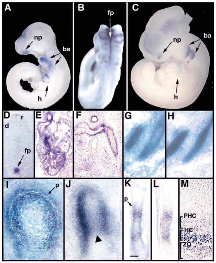
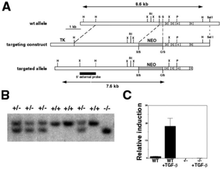
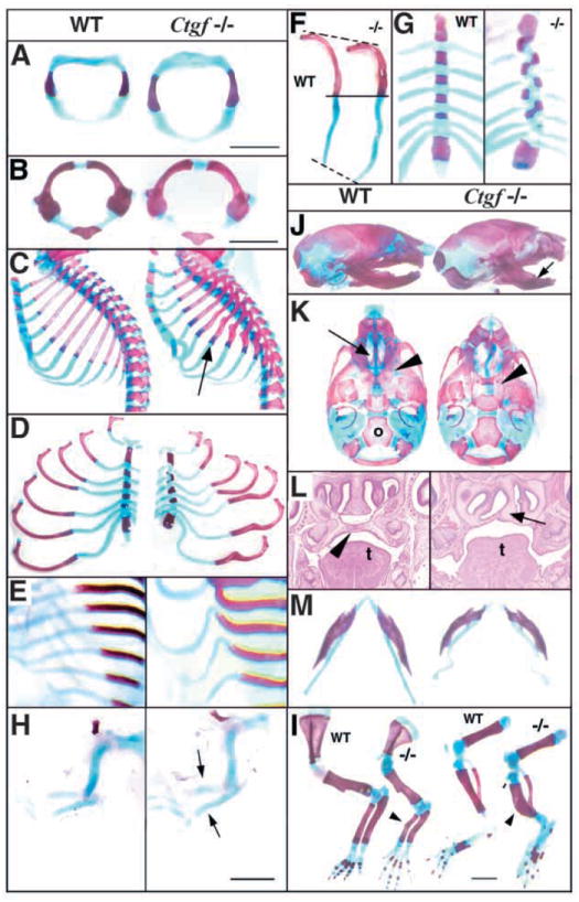
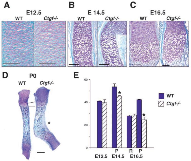
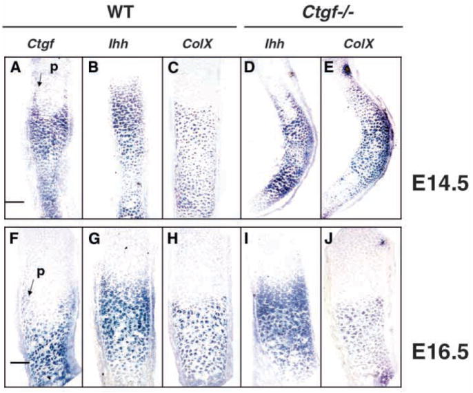
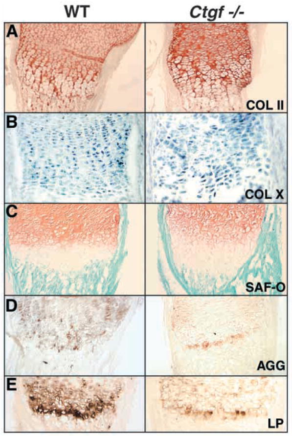
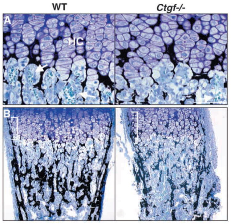
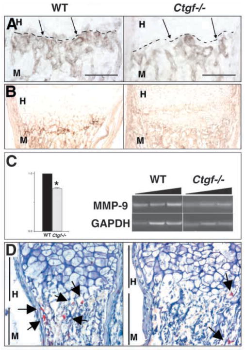
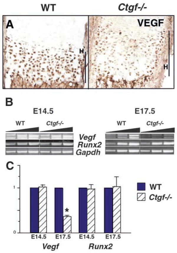
Coordinated production and remodeling of the extracellular matrix is essential during development. It is of particular importance for skeletogenesis, as the ability of cartilage and bone to provide structural support is determined by the composition and organization of the extracellular matrix. Connective tissue growth factor (CTGF, CCN2) is a secreted protein containing several domains that mediate interactions with growth factors, integrins and extracellular matrix components. A role for CTGF in extracellular matrix production is suggested by its ability to mediate collagen deposition during wound healing. CTGF also induces neovascularization in vitro, suggesting a role in angiogenesis in vivo. To test whether CTGF is required for extracellular matrix remodeling and/or angiogenesis during development, we examined the pattern of Ctgf expression and generated Ctgf-deficient mice. Ctgf is expressed in a variety of tissues in midgestation embryos, with highest levels in vascular tissues and maturing chondrocytes. We confirmed that CTGF is a crucial regulator of cartilage extracellular matrix remodeling by generating Ctgf(-/-) mice. Ctgf deficiency leads to skeletal dysmorphisms as a result of impaired chondrocyte proliferation and extracellular matrix composition within the hypertrophic zone. Decreased expression of specific extracellular matrix components and matrix metalloproteinases suggests that matrix remodeling within the hypertrophic zones in Ctgf mutants is defective. The mutant phenotype also revealed a role for Ctgf in growth plate angiogenesis. Hypertrophic zones of Ctgf mutant growth plates are expanded, and endochondral ossification is impaired. These defects are linked to decreased expression of vascular endothelial growth factor (VEGF) in the hypertrophic zones of Ctgf mutants. These results demonstrate that CTGF is important for cell proliferation and matrix remodeling during chondrogenesis, and is a key regulator coupling extracellular matrix remodeling to angiogenesis at the growth plate.


Figures

Fig. 1
Fig. 1
Expression of Ctgf in midgestation embryos. Whole-mount in situ hybridization of E9.5 (A,B) and an E10.5 (C) embryo treated with antisense Ctgf, demonstrating expression in heart, branchial arches, neuronal tissues and nasal process. (D) Frontal section through an E11.5 embryo showing high levels of Ctgf expression in the floorplate, and lower levels in the roofplate and dermomyotome. (E) Ctgf expression in the heart at E13.5. Transcripts are present in ventricular myofibroblasts and in fusing endocardial cushion tissue at the midline of the outflow tract. (F) Ctgf expression in the endothelium and smooth muscle layers of major blood vessels at E13.5. (G,H) Adjacent parasagittal sections through E12.5 ribs showing Ctgf expression in perichondrium (G) and collagen type II (ColII) (H) expression in proliferating chondrocytes. (I) Transverse section through an E13.5 femur showing Ctgf expression in perichondrium and in adjacent chondrocytes. (J) Sagittal section through the femur of an E13.5 embryo showing strong Ctgf expression in the perichondrium, and lower levels in chondrocytes at the center of the diaphysis (arrowhead). (K-M) Adjacent sections though an E14.5 femur showing expression of Ctgf (K), and Ihh (L). Scale bar in K: 100 μm for K,L. Ctgf is expressed in the domain of Ihh expression, as well as in prehypertrophic chondrocytes adjacent to the perichondrium. (M) Section through the proximal growth plate of a P0 femur, showing Ctgf expression in hypertrophic chondrocytes. ba, branchial arch; d, dermomyotome; fp, floorplate; h, heart; HC, hypertrophic chondrocytes; m, meninges; np, nasal process; p, perichondrium; PHC, prehypertrophic chondrocytes; r, roofplate; sc, spinal cord; ZO, zone of ossification.
Fig. 2
Fig. 2
Generation of Ctgf−/− mice. (A) Map of the Ctgf locus (wild-type allele), targeting vector and mutant allele produced by homologous recombination. The HindIII fragments expected for the wild-type (8.6 kb) and mutant (7.6 kb) alleles are indicated by double-headed arrows. A, ApaI; B, BamHI; C, ClaI; H, HindIII; N, NotI; P, PstI; R, EcoRI; S, SmaI; X, XbaI. (B) Southern blot analysis of genomic DNA from a heterozygote intercross. (C) Verification that the targeted locus encodes a null allele. RT-PCR analysis of fibroblasts from wild-type and mutant embryos treated with TGFβ1 (5 ng/ml) to induce Ctgf expression. No Ctgf transcripts can be detected in mutants, even after TGFβ treatment.
Fig. 3
Fig. 3
Ctgf−/− mice exhibit multiple skeletal defects. In all panels, wild-type is towards the left and Ctgf−/−is to the right. Atlases from E14.5 (A) and P0 (B) mutants are broader than those from wild-type littermates. (C) Sagittal views of neonatal rib cages showing deformation of cartilage, and kinks in bone in Ctgf−/− mice (arrow). (D) Flat mounts of rib cages show that in Ctgf mutants, ribs are kinked and the sternum is shortened. (E) E14.5 rib cages, showing that the kinks seen in neonatal mutant ribs are preceded by distorted rib cartilage. (F) Endochondral ossification is delayed in Ctgf mutants. Seventh ribs from neonatal wild-type and mutant littermates are shown. (G) Misalignment of the sternal bars is seen in ~10% of neonatal mutants. (H) Cleared skeletal preparations of E13.5 forelimbs, showing kinks in the radius and ulna (arrows) prior to ossification. (I) Cleared skeletal preparations of neonatal forelimbs and hindlimbs showing deformations (arrowheads) in the radius and ulna, and tibia and fibula. (J) Side views of neonatal skulls showing domed skull and shortened mandibles (arrow). (K) Ventral views of neonatal skulls, showing lack of elevation of the palatal shelves, leading to cleft palate (arrowhead), deformation of nasal cartilage, and consequent absence of the adjacent ethmoid bones (arrow in wild type), but no apparent defects in other membrane bones such as the occipital (o). (L) Frontal sections of E15.5 skulls, showing cleft palate (palate in wild type is indicated by arrowhead) and deformed nasal cartilage (arrow in mutant) in mutants. t, tongue. (M) Ctgf mutant neonates exhibit deformation of Meckel’s cartilage and shortened mandibles. Scale bars: 1 mm.
Fig. 4
Fig. 4
Histological and proliferative defects in Ctgf mutant cartilage. (A) Sections through wild-type and Ctgf mutant humeri at E12.5, showing no apparent differences in size or morphology. Scale bar: 50 μm. (B) Sections through wild-type and Ctgf mutant E14.5 radii at the metaphysis. Hypertrophic cells are present in wild-type and mutant littermates. Scale bar: 100 μm. (C) Sections through growth plates of E16.5 wild-type and Ctgf mutant radii demonstrate that the growth plate is expanded in mutants. The junction between the zones occupied by prehypertrophic and hypertrophic chondrocytes is disorganized in mutants. Scale bar: 100 μm. (D) Radii from newborn wild-type and Ctgf mutant littermates, demonstrating persistence of the enlarged hypertrophic zone. The epiphyses appear normal in mutants. The concave surface of the kink in mutants is a site of membrane bone formation (asterisk). Scale bar: 300 μm. (E) Reduced rates of chondrocyte cell proliferation in Ctgf mutants. Quantification of PCNA labeling is shown in the graph, with values expressed as % labeled nuclei. Cells in five adjacent sections, each spanning 40 μm, were scored by an observer blinded to the genotype. Statistical significance was assessed by Student’s t-test. *P<0.01; P, proliferative zone; R, resting zone.
Fig. 5
Fig. 5
Expression of Ihh and ColX in Ctgf mutants. (A-C) Expression of Ctgf (A), Ihh (B) and ColX (C) in the radius of an E14.5 wild-type embryo. The expression patterns of Ctgf and Ihh overlap in prehypertrophic and hypertrophic chondrocytes. Ctgf is co-expressed with ColX in hypertrophic chondrocytes at this stage and in prehypertrophic chondrocytes adjacent to the perichondirum (arrow in A). (D,E) Expression of Ihh (D) and ColX (E) in the radius of an E14.5 Ctgf mutant littermate. These markers are expressed in appropriate patterns in prehypertrophic and hypertrophic chondrocytes. Scale bar: 100 μm for A-E (F-H) Expression of Ctgf (F), Ihh (G) and ColX (I) in the radius of an E16.5 wild-type embryo. Ctgf is expressed primarily in hypertrophic chondrocytes in a pattern overlapping that of ColX, but transcripts also persist in chondrocytes adjacent to the perichondrium in the prehypertrophic zone (arrow in F). (I,J) Expression of Ihh (I) and ColX (J) in the radius of an E16.5 Ctgf mutant littermate. The domains of expression of Ihh are indistinguishable in wild-type and mutant littermates, indicating that the expansion of the hypertrophic zone revealed by histological analysis is not accompanied by an expanded prehypertrophic zone. Scale bar: 50 μm for F-J.
Fig. 6
Fig. 6
Expression of ECM components is altered in Ctgf mutants. (A) Immunostaining for type II collagen protein is at comparable intensities in P0 growth plates of Ctgf mutants and wild-type littermates. (B) Levels of type X collagen mRNA are indistinguishable in wild-type and Ctgf mutant growth plates. (C) Safranin-o staining demonstrates that proteoglycan levels are normal in the resting and proliferative zones, and that the hypertrophic zone, which is not stained intensely by safranin-O, is expanded in mutants. (D,E) Expression of aggrecan (D) and link protein (E) is reduced in P0 growth plates of Ctgf mutants.
Fig. 7
Fig. 7
Impaired angiogenesis and osteopenia in growth plates of Ctgf mutants. (A) Plastic sections through the growth plates of P0 femora stained by the method of von Kossa. The ECM of Ctgf mutants is mineralized (black stain), but hypertrophic chondrocyte columns (HC) are disorganized, and there are few capillaries (arrows) invading the cartilage matrix. A single capillary can be seen in the vicinity of the mutant growth plate. (B) von Kossa-stained plastic sections through neonatal femora from wild-type and Ctgf−/−mice demonstrate that mutants are osteopenic; the amount of mineralization (black stain) is greatly reduced in mutants. The bone collar (brackets) adjacent to the expanded hypertrophic zone is lengthened in mutants, but is thinner than in wild-type littermates. Scale bars: 40 μm.
Fig. 8
Fig. 8
Defective expression of angiogenic factors in Ctgf mutant growth plates. Growth plates of neonatal radii are shown in all panels. (A) PECAM immunostaining demonstrates the existence of blood vessels in the intertrabecular spaces of the metaphysis, but the fine network of capillaries seen at the ossification zone (broken line) in wild-type animals is not as extensive in Ctgf mutants. Scale bar: 100 μm. (B) MMP9 immunostaining is reduced in neonatal mutants. (C) Mmp9 RNA levels are reduced in neonatal mutants. The triangular bars above the photograph of the gel represent increasing numbers of amplification cycles (see Materials and Methods). Data are from three pairs on Ctgf mutants and wild-type littermates. Expression of Mmp9 was normalized to Gapdh in each reaction. *P<0.05. (D) TRAP-positive cells (arrows) are present in normal levels in the bone marrow of Ctgf mutants, but in reduced levels at the cartilage-bone junction, indicating a defect in recruitment of osteoclasts/chondroclasts to the hypertrophic region. MMP13 protein is present at diminished levels in hypertrophic chondrocytes in Ctgf mutants. H, hypertrophic zone; M, metaphysis.
Fig. 9
Fig. 9
VEGF expression is reduced in the hypertrophic zones of Ctgf mutant growth plates. (A) VEGF immunostaining is reduced in the hypertrophic zones of newborn Ctgf mutants. (B) Semi-quantitative RT-PCR analysis of Vegf and Runx2 expression. Representative data from a single Ctgf mutant and wild-type littermate at E14.5 and at E17.5 is shown. The triangular bars above the photograph of the gel represent increasing numbers of amplification cycles (see Materials and Methods). Expression of Runx2 and Vegf was normalized to Gapdh in each reaction. (C) Expression of Runx2 and Vegf in Ctgf mutants relative to wild-type littermates. Data are from five pairs of Ctgf mutants and wild-type littermates. Expression of Runx2 is not altered in mutants at E14.5 or E17.5. Vegf transcripts are present at indistinguishable levels in Ctgf mutants and wild-type littermates at E14.5, but at reduced levels in mutants at E17.5; the upper and lower bands correspond to the 165 and 120 Vegf isoforms, respectively. * P<0.05. H, hypertrophic zone.

References

    1. Abbondanzo S, Gadi I, Stewart C. Derivation of embryonic stem cell lines. Methods Enzymol. 1993;225:803–823. - PubMed
    1. Abreu G, Ketpura N, Reversade B, De Robertis E. Connective-tissue growth factor (CTGF) modulates cell signalling by BMP and TGF-β. Nat Cell Biol. 2002;4:599–604. - PMC - PubMed
    1. Ayer-Lelievre C, Brigstock D, Lau L, Pennica D, Perbal B, Yeger H. Report and abstracts on the first international workshop on the CCN family of genes. Mol Pathol. 2001;54:105–120. - PMC - PubMed
    1. Babic AM, Chen C-C, Lau L. Fisp12/mouse connective tissue growth factor mediates endothelial cell adhesion and migration through integrin αvβ3, promotes endothelial cell survival, and induces angiogenesis in vivo. Mol Cell Biol. 1999;19:2958–2966. - PMC - PubMed
    1. Bitgood MJ, McMahon AP. Hedgehog and Bmp genes are coexpressed at many diverse sites of cell-cell interaction in the mouse embryo. Dev Biol. 1995;172:126–138. - PubMed

Publication types

MeSH terms

Substances