. 2003 May 15;31(10):2694-702.
doi: 10.1093/nar/gkg364.

The (52-96) C-terminal domain of Vpr stimulates HIV-1 IN-mediated homologous strand transfer of mini-viral DNA

Affiliations

The (52-96) C-terminal domain of Vpr stimulates HIV-1 IN-mediated homologous strand transfer of mini-viral DNA

Julien Bischerour et al. Nucleic Acids Res. .

Abstract

Viral integrase (IN) and Vpr are both components of the human immunodeficiency virus type 1 (HIV-1) pre-integration complex. To investigate whether these proteins interact within this complex, we investigated the effects of Vpr and its subdomains on IN activity in vitro. When a 21mer oligonucleotide was used as a donor and acceptor, both Vpr and its C-terminal DNA-binding domain [(52-96)Vpr] inhibited the integration reaction, whereas the (1-51)Vpr domain did not affect IN activity. Steady-state fluorescence anisotropy showed that both full-length and (52-96)Vpr bind to the short oligonucleotide, thereby extending previous observations with long DNA. The concentrations of the two proteins required to inhibit IN activity were consistent with their affinities for the oligonucleotide. The use of a 492 bp mini-viral substrate confirmed that Vpr can inhibit the IN-mediated reaction. However, the activity of (52-96)Vpr differed notably since it stimulated specifically integration events involving two homologous mini-viral DNAs. Order of addition experiments indicated that the stimulation was maximal when IN, (50-96)Vpr and the mini-viral DNA were allowed to form a complex. Furthermore, in the presence of (50-96)Vpr, the binding of IN to the mini-viral DNA was dramatically enhanced. Taken together, these data suggest that (52-96)Vpr stimulates the formation of a specific complex between IN and the mini-viral DNA.


Figures
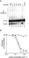
Figure 1
Figure 1
Inhibition of HIV-1 IN-mediated 3′ processing by Vpr and its subdomains. (A) Dose–response effect of full-length Vpr on 3′ processing of a 21mer ODN substrate. The 5′-end-labeled U5 substrate (10 nM) was incubated for 1 h at 37°C with 65 nM IN. Lanes 1–6, 50, 100, 200, 400 and 800 nM, and 1.6 µM Vpr, respectively; lane 7, no Vpr; lane 8, negative control in the absence of IN. The 21mer DNA substrate, 19mer 3′ processed product and the strand transfer products are indicated on the left. (B) Quantification of experimental data obtained in the presence of full-length Vpr (filled circles), (1–51) (open squares) and (52–96) (open diamonds) peptides of Vpr. Data from two independent experiments were averaged.
Figure 2
Figure 2
Binding of Vpr and (52–96)Vpr to the double-stranded 21mer U5 ODN. Change in steady-state fluorescence anisotropy of fluorescein-labeled U5 ODN (10 nM) was monitored as a function of increasing concentrations of either full-length Vpr (filled squares) or (52–96)Vpr (filled triangles). The anisotropy value of labeled dsDNA alone recorded in our condition was close to 0.04 and was systematically subtracted from the anisotropy value of the DNA–protein complex (δ anisotropy). Addition of (1–51)Vpr did not lead to a change in anisotropy, indicating the absence of complex formation. Isotherm binding curves were fitted with Prism3.0 software.
Figure 3
Figure 3
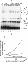
Vpr effects on long DNA integration activity. (A) Phosphorimage of long DNA strand transfer assay. Reactions were performed by incubation for 1 h at 37°C of 2 pmol IN, 10 ng of U3U5 substrate and 40 ng of pSP70 target in the presence of increasing concentrations of Vpr and its subdomains: 0.4 µM (lanes 3, 6 and 9), 0.8 µM (lanes 4, 7 and 10), 1.6 µM (lanes 5, 8 and 11) of Vpr, (1–51)Vpr or (52–96)Vpr, respectively. Components were pre-incubated for 10 min on ice before incubation at 37°C, with the exception of the pSP70 target, which was added at the beginning of the reaction. The nature of the integration product is reported on the right: bold arrows indicate the main products corresponding to integration of one mini-viral DNA; fainter arrows indicate products corresponding to multiple integration events. (a) 32P-labeled U3U5 substrate; (b) homologous integration products; (c) heterologous integration products. (B) Quantification of homologous integration products using ImageQuant software. Homologous integration in the presence of (52–96)Vpr (filled circles); (1–51)Vpr (filled inverted triangles) and Vpr (filled squares).
Figure 4
Figure 4
Effects of the overlapping C-terminal peptides of Vpr on mini-viral DNA strand transfer activity. Reactions were performed by incubation for 1 h at 37°C of 2 pmol IN, 10 ng of U3U5 substrate and 40 ng of pSP70 target in the presence of increasing concentrations of Vpr peptides. Concentrations of 0.8, 1.6, 3.2 µM of (52–96) were used in lanes 3–5, of (52–70) in lanes 6–8, of (70–96) in lanes 9–11 and of (60–80) in lanes 12–14, without Vpr peptides in lane 2 or without IN in lane 1. Components were pre-incubated together for 10 min on ice before incubation at 37°C. (A) Phosphorimage of heterologous strand transfer; bands located above the main products correspond to multiple integration events. (B) Phosphorimage of homologous strand transfer products.
Figure 5
Figure 5
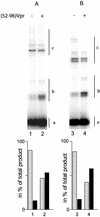
Effect of (52–96) peptide on mini-viral DNA strand transfer using a linear target. The reaction was performed as described in Figure 3, except that the circular pSP70 plasmid was exchanged with either the circular pU3U5 plasmid DNA (A) or with a linear 2500 bp target resulting from the digestion of the pU3U5 plasmid with NdeI (B). The assay was carried out in the absence (lanes 1 and 3) or presence (lanes 2 and 4) of 0.8 µM of (52–96)Vpr. The nature of the integration product is reported on the right: 32P-labeled U3U5 substrate (a), homologous integration products (b) and heterologous integration products (c).
Figure 6
Figure 6
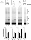
Effect of the pre-incubation of the partners on the (52–96)Vpr stimulation of the long DNA strand transfer. Reactions were performed with 2 pmol IN, 10 ng of U3U5 substrate and 40 ng of pSP70 target in the presence (lanes 2, 4, 6 and 7) or absence (lanes 1, 3 and 5) of (52–96)Vpr. Either all the partners are pre-incubated for 10 min on ice before 1 h of incubation at 37°C (lanes 1 and 2), U3U5 DNA donor is omitted from the pre-incubation (lanes 3 and 4) or the pSP70 target is omitted from the pre-incubation (lanes 5 and 6). Finally, IN and U3U5 on one side and (52–96)Vpr with pSP70 on the other side were pre-incubated for 10 min on ice before being mixed together (lane 7). The nature of the integration product is reported on the right: 32P-labeled U3U5 substrate (a), homologous integration products (b) and heterologous integration products (c).
Figure 7
Figure 7
Electrophoretic mobility gel shift assay with IN and (52–96) peptide. Incubations were performed for 30 min on ice with 10 ng of radiolabeled U3U5 substrate and 40 ng of pSP70 target in a buffer containing 20 mM HEPES pH 7.0, 2 mM DTT and 10 mM MnCl2. Gel shift assay with increasing concentrations of IN: 0.1, 0.2, 0.4 and 0.6 µM (lanes 2–5). Lane 1, no IN (left panel). Gel shift assay with increasing concentrations of (52–96)Vpr: 0.2, 0.4, 0.8 and 1.6 µM (lanes 7–10). Lane 6, no (52–96)Vpr (center panel). Gel shift assay with increasing concentrations of (52–96)Vpr in the presence of IN at the concentration of 0.2 µM (right panel). The concentrations of (52–96)Vpr were identical to those in the center panel. Asterisks indicate discrete, stable complexes which were observed only in the concomitant presence of IN and (52–96)Vpr.

Similar articles

Cited by

References

    1. Brown P.O., Bowerman,B., Varmus,H.E. and Bishop,J.M. (1989) Retroviral integration: structure of the initial covalent product and its precursor and a role for the viral IN protein. Proc. Natl Acad. Sci. USA, 86, 2525–2529. - PMC - PubMed
    1. Fujiwara T. and Mizuuchi,K. (1988) Retroviral DNA integration: structure of an integration intermediate. Cell, 54, 497–504. - PubMed
    1. Carteau S., Gorelick,R.J. and Bushman,F.D. (1999) Coupled integration of human immunodeficiency virus type 1 cDNA ends by purified integrase in vitro: stimulation by the viral nucleocapsid protein. J. Virol., 73, 6670–6679. - PMC - PubMed
    1. Cherepanov P., Surratt,D., Toelen,J., Pluymers,W., Griffith,J., De Clercq,E. and Debyser,Z. (1999) Activity of recombinant HIV-1 integrase on mini-HIV DNA. Nucleic Acids Res., 27, 2202–2210. - PMC - PubMed
    1. Hindmarsh P., Ridky,T., Reeves,R., Andrake,M., Skalka,A.M. and Leis,J. (1999) HMG protein family members stimulate human immunodeficiency virus type 1 and avian sarcoma virus concerted DNA integration in vitro. J. Virol., 73, 2994–3003. - PMC - PubMed

Publication types

MeSH terms