

Real-time PCR-based method for the estimation of genome sizes
- PMID: 12736322
- PMCID: PMC156059
- DOI: 10.1093/nar/gng056
Real-time PCR-based method for the estimation of genome sizes
Abstract
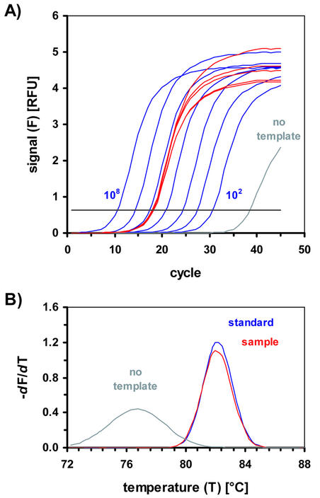
The fast and reliable estimation of the genome sizes of various species would allow for a systematic analysis of many organisms and could reveal insights into evolutionary processes. Many methods for the estimation of genome sizes have already been described. The classical methods are based on the determination of the phosphate content in the DNA backbone of total DNA isolated from a defined number of cells or on reassociation kinetics of high molecular weight genomic DNA (c(0)t assay). More recent techniques employ DNA-specific fluorescent dyes in flow cytometry analysis, image analysis or absorption cytometry after Feulgen staining. The method presented here is based on the absolute quantification of genetic elements in a known amount (mass) of genomic DNA by real-time quantitative PCR. The method was evaluated on three different eukaryotic species, Saccharomyces cerevisiae (12.1 Mb), Xiphophorus maculatus (550 Mb) and Homo sapiens sapiens (2.9 Gb), and found to be fast, highly accurate and reliable.
Figures
Similar articles
-
[Use of the real-time RT-PCR method for investigation of small stable RNA expression level in human epidermoid carcinoma cells A431].Tsitologiia. 2003;45(4):392-402. Tsitologiia. 2003. PMID: 14520871 Russian.
-
Developmental validation of a real-time PCR assay for the simultaneous quantification of total human and male DNA.Forensic Sci Int Genet. 2008 Dec;3(1):14-21. doi: 10.1016/j.fsigen.2008.07.004. Epub 2008 Sep 9. Forensic Sci Int Genet. 2008. PMID: 19083861
-
An event-specific method for the detection and quantification of ML01, a genetically modified Saccharomyces cerevisiae wine strain, using quantitative PCR.Int J Food Microbiol. 2016 Oct 3;234:15-23. doi: 10.1016/j.ijfoodmicro.2016.06.017. Epub 2016 Jun 20. Int J Food Microbiol. 2016. PMID: 27367966
-
[Quantitative PCR in the diagnosis of Leishmania].Parassitologia. 2004 Jun;46(1-2):163-7. Parassitologia. 2004. PMID: 15305709 Review. Italian.
-
Rapid amplification of uncharacterized transposon-tagged DNA sequences from genomic DNA.Yeast. 1997 Mar 15;13(3):233-40. doi: 10.1002/(SICI)1097-0061(19970315)13:3<233::AID-YEA88>3.0.CO;2-E. Yeast. 1997. PMID: 9090052
Cited by
-
Comparative genomic analysis of the 'pseudofungus' Hyphochytrium catenoides.Open Biol. 2018 Jan;8(1):170184. doi: 10.1098/rsob.170184. Open Biol. 2018. PMID: 29321239 Free PMC article.
-
Intraspecific variation in genome size in Artemisia argyi determined using flow cytometry and a genome survey.3 Biotech. 2023 Feb;13(2):57. doi: 10.1007/s13205-022-03412-y. Epub 2023 Jan 22. 3 Biotech. 2023. PMID: 36698769 Free PMC article.
-
A draft genome sequence of an invasive mosquito: an Italian Aedes albopictus.Pathog Glob Health. 2015 Jul;109(5):207-20. doi: 10.1179/2047773215Y.0000000031. Epub 2015 Sep 14. Pathog Glob Health. 2015. PMID: 26369436 Free PMC article.
-
Transcriptome and genome size analysis of the Venus flytrap.PLoS One. 2015 Apr 17;10(4):e0123887. doi: 10.1371/journal.pone.0123887. eCollection 2015. PLoS One. 2015. PMID: 25886597 Free PMC article.
-
Genomic and transcriptomic comparison of nucleotide variations for insights into bruchid resistance of mungbean (Vigna radiata [L.] R. Wilczek).BMC Plant Biol. 2016 Feb 17;16:46. doi: 10.1186/s12870-016-0736-1. BMC Plant Biol. 2016. PMID: 26887961 Free PMC article.
References
-
- Zhang J.Z and Fan,M.Y. (2002) Determination of genome size and restriction fragment length polymorphism of four Chinese rickettsial isolates by pulsed-field gel electrophoresis. Acta Virol., 46, 25–30. - PubMed
-
- Rydkina E., Roux,V. and Raoult,D. (1999) Determination of the genome size of Ehrlichia spp., using pulsed field gel electrophoresis. FEMS Microbiol. Lett., 176, 73–78. - PubMed
-
- Shapiro H.S. (1970) Nucleic Acids. In Sober,H.A. (ed.), CRC Handbook of Biochemistry. Selected Data for Molecular Biology. CRC Press, Cleveland, OH, pp. H-113.
-
- Britten R.J. and Kohne,D.E. (1986) Repeated sequences in DNA. Hundreds and thousands of copies of DNA sequences have been incorporated into the genomes of higher organisms. Science, 161, 529–540. - PubMed
Publication types
MeSH terms
Substances
LinkOut - more resources
Full Text Sources
Molecular Biology Databases
Miscellaneous
