. 2003 May 1;23(9):3597-606.
doi: 10.1523/JNEUROSCI.23-09-03597.2003.

Sp1 and Sp3 are oxidative stress-inducible, antideath transcription factors in cortical neurons

Affiliations

Sp1 and Sp3 are oxidative stress-inducible, antideath transcription factors in cortical neurons

Hoon Ryu et al. J Neurosci. .

Abstract

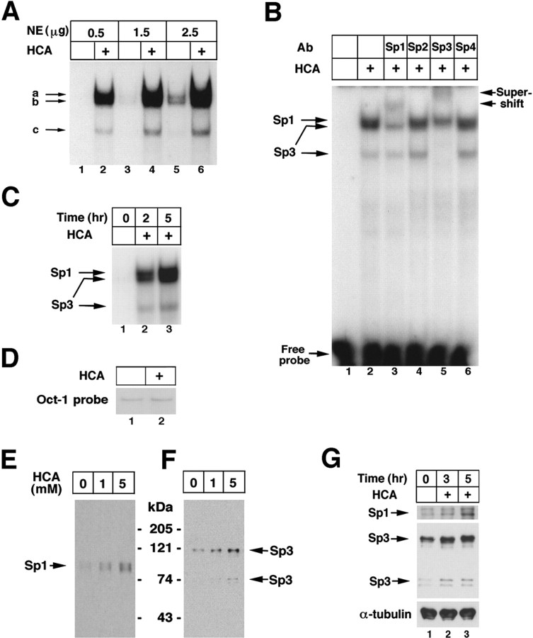
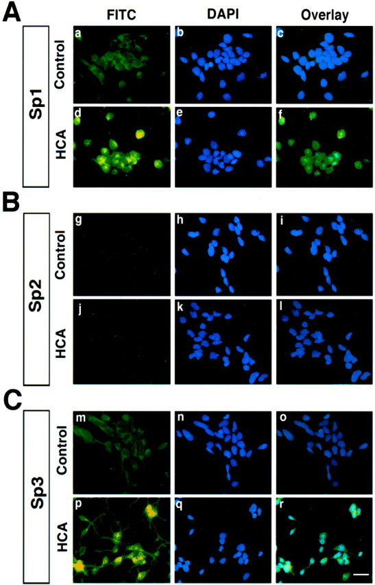
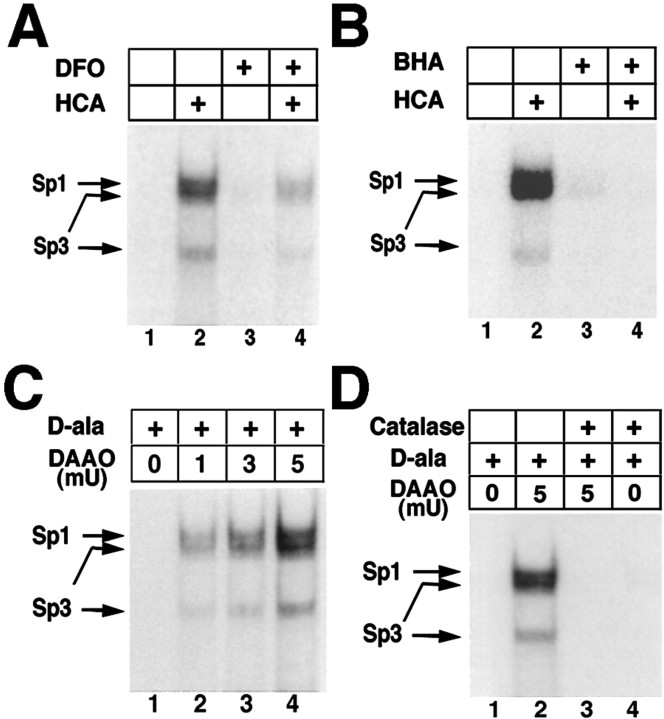
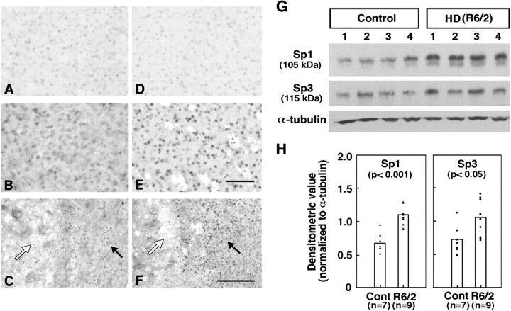
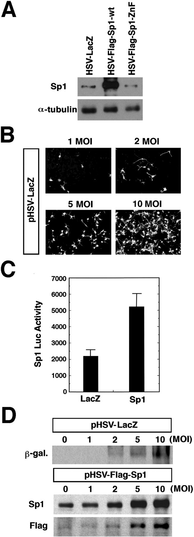
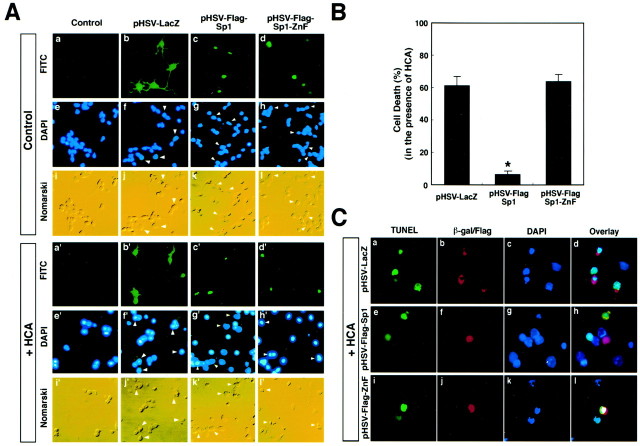
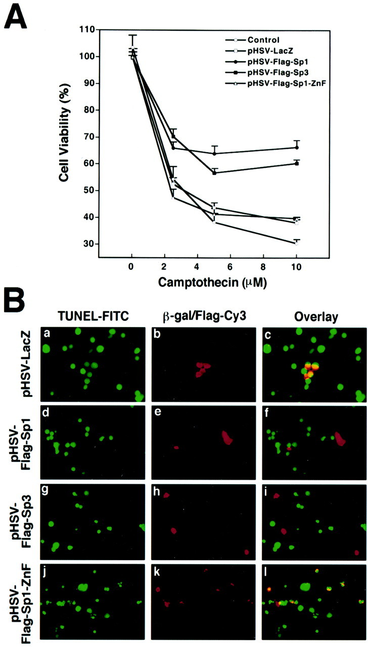
Neuronal cell death in response to oxidative stress may reflect the failure of endogenous adaptive mechanisms. However, the transcriptional activators induced by oxidative stress in neurons that trigger adaptive genetic responses have yet to be fully elucidated. We report that basal DNA binding of the zinc finger transcription factors Sp1 and Sp3 is unexpectedly low in cortical neurons in vitro and is significantly induced by glutathione depletion-induced or hydrogen peroxide-induced oxidative stress in these cells. The increases in Sp1/Sp3 DNA binding reflect, in part, increased levels of Sp1 and Sp3 protein in the nuclei of cortical neurons. Similar induction of Sp1 and Sp3 protein is also observed in neurons in vivo in a chemical or a genetic model of Huntington's disease, two rodent models in which neuronal loss has been attributed to oxidative stress. Sustained high-level expression of full-length Sp1 or full-length Sp3, but not the Sp1 zinc finger DNA-binding domain alone, prevents death in response to oxidative stress, DNA damage, or both. Taken together, these results establish Sp1 and Sp3 as oxidative stress-induced transcription factors in cortical neurons that positively regulate neuronal survival.


Figures

Fig. 1.
Fig. 1.
Glutathione depletion-induced oxidative stress induces Sp1 and Sp3 DNA-binding activity in cortical neurons.A, EMSA performed with increasing amounts of protein from nuclear extracts (NEs) from control (lanes 1, 3, 5) and HCA (a glutamate analog)-treated neurons (lanes 2, 4, 6), 4 hr after the onset of HCA treatment. The three DNA-binding activities induced by oxidative stress are designated a–c. B,Identification of Sp1 in complex a and Sp3 in complexesb and c by supershift analysis using subunit-specific antibodies to Sp1–Sp4. C, Time course of induction of Sp1 and Sp3 DNA-binding activities after HCA (1 mm) treatment. D, The effect of HCA-induced glutathione depletion on Oct-1 DNA-binding activity is shown 4 hr after the onset of HCA treatment. Effects of increasing concentrations of HCA on nuclear Sp1 (E) and Sp3 (F) protein levels 4–5 hr after the onset of HCA treatment are shown. G, Time course of changes of nuclear Sp1 and Sp3 levels after the onset of HCA treatment. Examples are representative of three to five independent experiments.
Fig. 2.
Fig. 2.
HCA-induced glutathione depletion and oxidative stress induce Sp1 (A) and Sp3 (C), but not Sp2 (B), in the nucleus of embryonic cortical neurons. Immunocytochemical analysis of Sp1 (a, d), Sp2 (i), Sp3 (m, p), and the nuclear stain DAPI (b, e, h, k, n, q) in mock-treated (a, g, m, b, h, n) or HCA-treated (d, j, e, k, q) mixed cortical neuronal cultures. Cells were plated on eight-well chamber slides for 24 hr and treated with HCA (3 mm) for 3 hr. Cells were then fixed with 4% PFA for 15 min, and immunofluorescence staining was performed as described in Materials and Methods. Scale bar, 20 μm.
Fig. 3.
Fig. 3.
Increases in Sp1 and Sp3 DNA binding induced by the glutamate analog HCA are inhibited by antioxidants; Sp1 and Sp3 DNA binding in cortical neurons are activated by hydrogen peroxide. Induction of Sp1 and Sp3 DNA binding by HCA-induced glutathione depletion (4 hr) is decreased by the antioxidant iron chelator DFO (100 μm;A) and the lipid peroxidation inhibitor BHA (10 μm;B).C, Addition of exogenous peroxide, generated by the enzyme DAAO and its substrate d-ala (20 mm) for 4 hr increases Sp1 and Sp3 DNA binding in a concentration-dependent manner in cortical neurons. The induction is observed despite no morphological or biochemical evidence of cell death in cortical neurons. D, Addition of catalase abrogates Sp1 and Sp3 DNA binding induced by d-ala (20 mm) and DAAO (5 mU). Examples are representative of three to five independent experiments.
Fig. 4.
Fig. 4.
Sp1 and Sp3 tissue immunoreactivity in R6/2 transgenic mice and 3-NP-lesioned-mice. Sp1 (A–C) and Sp3 (D–F) immunoreactivities in the neostriatum of 12-week-old wild-type littermate control mice (A, D) and R6/2 transgenic HD mice (B, E) and 3-NP-lesioned wild-type mice (C, F) are shown. Sp1 and Sp3 immunostaining of neurons in wild-type control mice (A, D) is shown. Markedly increased Sp1 and Sp3 immunoreactivity was observed in R6/2 mice compared with wild-type controls, with the greatest increases in Sp3 immunoreactivity. In 3-NP-lesioned mice, Sp1 (C) and Sp3 (F) immunoreactivities were both increased within neurons surrounding the lesion core (asterisk) within the penumbra or transition zone of neuronal injury. Scale bars: (in E) A, B,D, E, 100 μm; (in F), C, F, 200 μm. G, Sp1 and Sp3 protein levels in 12-week-old littermate controls and R6/2 transgenic mouse model of HD. α-Tubulin was used as a loading control. H, Scatter plot of densitometric values (normalized to α-tubulin) for Sp1 and Sp3 protein levels in seven littermate control and nine R6/2 HD mice.
Fig. 5.
Fig. 5.
HSV can be used to achieve expression of heterologous Sp1 in cortical neurons. A, Immunoblot demonstrating expression of heterologous Sp1 in cortical neurons 24 hr after infection with HSV vectors. HSV has an MOI of 2.B, HSV-LacZ leads to MOI-dependent β-galactosidase expression in cortical neurons. C, HSV-Sp1 infection of cortical neurons leads to induction of luciferase activity driven by Sp1 response elements compared with HSV-LacZ. Results are means ± SE for three separate experiments. D, Immunoblot analysis confirms MOI-dependent, HSV vector-driven expression of Sp1 (HSV-Sp1) or β-galactosidase. In parallel, immunoblotting with Flag antibody confirms that HSV-Sp1 results in heterologous rather than endogenous Sp1 expression.
Fig. 6.
Fig. 6.
Overexpression of Sp1 (HSV-Flag-Sp1), but not the Sp1 Zn finger DNA binding domain alone (HSV-Flag-Sp1-ZnF) or HSV-LacZ, in cortical neurons inhibits oxidative stress-induced cell death in cortical neurons. A, DAPI staining of LacZ-positive, Flag-Sp1-positive, or Flag-Sp1-ZnF-positive cortical neurons. The FITC column reflects FITC secondary antibody that recognizes LacZ-, Flag-Sp1-wt-, or Flag-Sp1-ZnF-expressing cortical neurons. Note the presence of LacZ in cell bodies as well as neurites, which is typical of cortical neurons in culture. Also note the presence of Sp1-wt and Sp1-ZnF in the nucleus, the expected localization for these transcription factors. The DAPI column shows the DAPI staining of FITC-positive cells in the same row, along with a few surrounding FITC-negative cells. Note that HCA produces an increase in the percentage of cells with pyknotic nuclei that display stronger fluorescence, which is characteristic of apoptotic cells. However, the Flag-Sp1-wt-expressing neurons display dim, diffuse DAPI staining, which is characteristic of normal cells. Cortical neurons were infected with 2–5 MOI of HSV vectors. B, Quantitative analysis. Overexpression of Flag-Sp1-wt but not Flag-Sp1-ZnF or LacZ blocks the increase in apoptotic cells elicited by HCA (LacZ or Sp1-ZnF vs Sp1-wt,p < 0.01). At least 300 cells were counted in each group, and the results are means ± SE for three different slides.C, TUNEL was performed to measure HCA-induced DNA damage of LacZ-positive, Flag-Sp1-positive, and Flag-Sp1-ZnF-positive cortical neurons. Cy3 (red) column reflects Cy3 secondary antibody that recognizes LacZ, Flag-Sp1, Flag-Sp3, or Sp1-ZnF. The overlay column reflects green TUNEL–FITC fluorescence superimposed on red Cy3 secondary antibody fluorescence for β-galactosidase, Sp1, and Sp1-ZnF and blue fluorescence for the nucleus.
Fig. 7.
Fig. 7.
Overexpression of Sp1-wt or Sp3-wt, but not the Sp1-ZnF or LacZ, inhibits neuronal death attributable to the DNA-damaging agent and topoisomerase inhibitor camptothecin.A, Quantitative analysis. Overexpression of Flag-Sp1 or Flag-Sp3 but not LacZ or Flag-Sp1-ZnF diminishes cell death induced by camptothecin (2–10 μm). Cortical neurons were infected with 2–5 MOI of HSV vectors. Cell viability was measured by incubating cortical neurons with MTT for 2 hr and measuring the amount of reduction to blue formazan. Results are means ± SE for three separate experiments. B, TUNEL, a marker of DNA double-strand breaks resulting from camptothecin-induced topoisomerase inhibition (Morris and Geller, 1996) of LacZ-positive, Flag-Sp1-positive, Flag-Sp3-positive, or Flag-Sp1-ZnF-positive cortical neurons. The Cy3 (red) column reflects Cy3 secondary antibody that recognizes LacZ, Flag-Sp1, Flag-Sp3, or Sp1-ZnF. The overlay column reflects green TUNEL–FITC fluorescence superimposed on red Cy3 secondary antibody fluorescence. Note that only cells expressing LacZ or Flag Sp1-ZnF are yellow, whereas Cy3-positive Flag-Sp1- and Flag-Sp3-expressing cells are not TUNEL labeled and are therefore red.

References

    1. Adida C, Crotty PL, McGrath J, Berrebi D, Diebold J, Altieri DC. Developmentally regulated expression of the novel cancer anti-apoptosis gene survivin in human and mouse differentiation. Am J Pathol. 1998;152:43–49. - PMC - PubMed
    1. Albers DS, Beal MF. Mitochondrial dysfunction and oxidative stress in aging and neurodegenerative disease. J Neural Transm [Suppl] 2000;59:133–154. - PubMed
    1. Atwood CS, Huang X, Moir RD, Tanzi RE, Bush AI. Role of free radicals and metal ions in the pathogenesis of Alzheimer's disease. Met Ions Biol Syst. 1999;36:309–364. - PubMed
    1. Beal MF, Brouillet E, Jenkins BG, Ferrante RJ, Kowall NW, Miller JM, Storey E, Srivastava R, Rosen BR, Hyman BT. Neurochemical and histological characterization of striatal excitotoxic lesions produced by the mitochondrial toxin, 3-nitropropionic acid. J Neurosci. 1993;13:4181–4192. - PMC - PubMed
    1. Beal MF. Oxidative metabolism. Ann NY Acad Sci. 2000;924:164–169. - PubMed

Publication types

MeSH terms