. 2003 May 1;23(9):3689-96.
doi: 10.1523/JNEUROSCI.23-09-03689.2003.

Heme oxygenase-2 protects against lipid peroxidation-mediated cell loss and impaired motor recovery after traumatic brain injury

Affiliations

Heme oxygenase-2 protects against lipid peroxidation-mediated cell loss and impaired motor recovery after traumatic brain injury

Edward F Chang et al. J Neurosci. .

Abstract

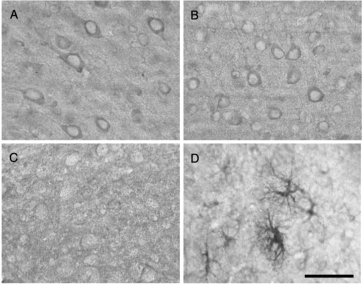
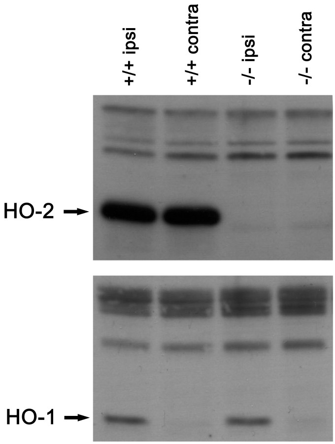
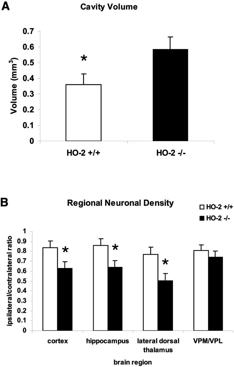
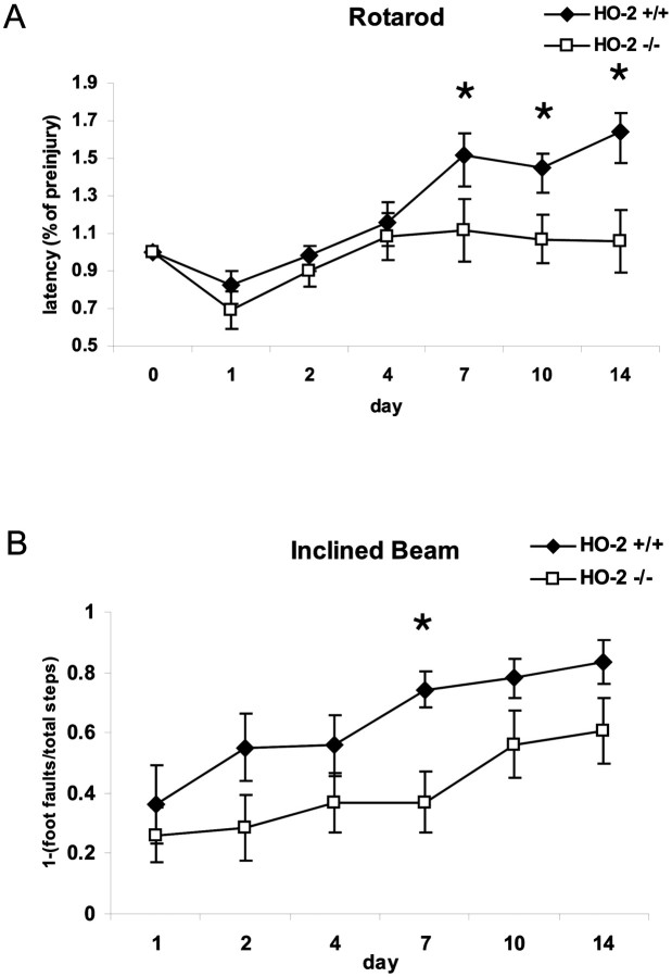
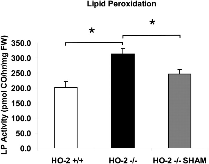
After traumatic brain injury (TBI), substantial extracellular heme is released from hemoproteins during hemorrhage and cell injury. Heme oxygenase (HO) isozymes are thought to detoxify the pro-oxidant heme to the potent antioxidant, bilirubin. HO-1, the inducible isozyme, is expressed in glial populations after injury and may play a protective role. However, the role of HO-2, the predominant and constitutively expressed isozyme in the brain, remains unclear after TBI. We used a controlled cortical impact injury model to determine the extent and mechanism of damage between HO-2 knock-out (KO) (-/-) and wild-type (WT) (+/+) mice. The specific cellular and temporal expressions of HO-2 and HO-1 were characterized by immunocytochemistry and Western blots. HO-2 was immunolocalized in neurons both before and after TBI, whereas HO-1 was highly upregulated in glia only after TBI. HO activity determined by gas chromatography using brain sonicates from injured HO-2 KO mice was significantly less than that of HO-2 wild types, despite the induction of HO-1 expression after TBI. Cell loss was significantly greater in KO mice in areas including the cortex, the CA3 region of hippocampus, and the lateral dorsal thalamus. Furthermore, motor recovery after injury, as measured by the rotarod assay and an inclined beam-walking task, was compromised in the KO mice. Finally, brain tissue from injured HO-2 KO mice exhibited decreased ability to reduce oxidative stress, as measured with an Fe(2+)/ascorbic acid-mediated carbon monoxide generation assay for lipid peroxidation susceptibility. These findings demonstrate that HO-2 expression protects neurons against TBI by reducing lipid peroxidation via the catabolism of free heme.


Figures

Fig. 1.
Fig. 1.
Immunolocalization of HO-2 and HO-1 in the uninjured and injured brain. HO-2 is constitutively expressed in cortical neurons in the uninjured brain (A) and after TBI (B) in the HO-2 wild-type mouse. HO-2 is localized primarily to neurons and is unchanged after injury. In contrast, HO-1 expression is virtually undetectable in the uninjured cortex (C) and highly induced in reactive astrocytes and microglia/macrophages after TBI (D). Scale bar, 50 μm.
Fig. 2.
Fig. 2.
Western blots from brain sonicates for HO-1 (bottom) and HO-2 (top) after TBI. Both ipsilateral and contralateral hemispheres are included. HO-1 is highly induced in the ipsilateral hemisphere, but only modestly induced in the contralateral hemisphere. In addition, HO-1 expression appears similar between hemispheres for both HO-2 wild-type and HO-2 KO mice. In contrast, strong HO-2 expression is found in both hemispheres of the HO-2 wild-type mice.
Fig. 3.
Fig. 3.
Total heme oxygenase (HO) activity by genotype. In uninjured mice (white bars), HO-2 wild-type (+/+) mice demonstrated the highest levels of HO activity. HO-2 heterozygous (+/−) mice showed relatively less activity, and HO-2 homozygous knock-out (−/−) mice showed the least activity (*p ≤ 0.05; ANOVA; Bonferroni t test). Activity in HO-2 KO mice is attributed primarily to the HO-1 isozyme. Total activity is increased at 24 hr after experimental injury in both HO-2 wild-type and HO-2 KO mice (black bars), suggesting induction of HO-1. Total activity, however, is still reduced significantly in HO-2 KO mice compared with HO-2 wild-type mice. Values are shown as the mean ± SEM (*p ≤ 0.05; unpaired t test).
Fig. 4.
Fig. 4.
Representative NeuN immunostained coronal section after TBI. At 14 d after injury, a large cortical cavitation at the impact site extends down to the level of the external capsule. Cell loss is discernible in the parenchyma immediately surrounding the cavity, ipsilateral hippocampal CA1 and CA3, and ipsilateral thalamic structures.
Fig. 5.
Fig. 5.
Cell loss after traumatic brain injury.A, Volumetric measurements of cortical cavity on histologic sections. Values are shown as the mean ± SEM (*p ≤ 0.05; unpaired t test).B, Regional neuronal density by NeuN cell counting in adjacent cortex, hippocampal CA3, lateral dorsal thalamus, and medial/lateral ventroposterior nuclei of thalamus. Values are shown as the mean ± SEM (*p ≤ 0.05; unpairedt test).
Fig. 6.
Fig. 6.
Motor recovery after experimental injury.A, The standard rotarod assay performance was expressed as the percentage of pretrauma latency. A repeated-measures ANOVA shows that overall rotarod performance is significantly worse in HO-2 KO mice compared with HO-2 wild-type mice. Post hoc analysis reveals that significant differences occur on days 7, 10, and 14. Values are shown as the mean ± SEM (*p ≤ 0.05; Bonferroni t test). B, Motor coordination was tested on an inclined beam walking task. Performance was expressed as the ratio of foot faults of the contralateral hindlimb to the number of total steps, subtracted from 1. Motor recovery is significantly worse in HO-2 KO compared with HO-2 wild-type mice (ANOVA), specifically on day 7 after injury. Values are shown as the mean ± SEM (*p ≤ 0.05; Bonferronit test).
Fig. 7.
Fig. 7.
Susceptibility to lipid peroxidation. An Fe2+- and ascorbate-containing in vitro lipid peroxidation assay measuring the generation of carbon monoxide (CO) was used to determine susceptibility of brain tissue to lipid peroxidation at 24 hr after experimental injury. Lipid peroxidation is higher in HO-2 KO mice compared with both HO-2 wild-type mice and sham-injured HO-2 KO mice. Values are shown as the mean ± SEM (*p ≤ 0.05; unpairedt test).

References

    1. Baranano DE, Snyder SH. Neural roles for heme oxygenase: contrasts to nitric oxide synthase. Proc Natl Acad Sci USA. 2001;98:10996–11002. - PMC - PubMed
    1. Baranano DE, Rao M, Ferris CD, Snyder SH. Biliverdin reductase: a major physiologic cytoprotectant. Proc Natl Acad Sci USA. 2002;99:16093–16098. - PMC - PubMed
    1. Burnett AL, Johns DG, Kriegsfeld LJ, Klein SL, Calvin DC, Demas GE, Schramm LP, Tonegawa S, Nelson RJ, Snyder SH, Poss KD. Ejaculatory abnormalities in mice with targeted disruption of the gene for heme oxygenase-2. Nat Med. 1998;4:84–87. - PubMed
    1. Conti AC, Raghupathi R, Trojanowski JQ, McIntosh TK. Experimental brain injury induces regionally distinct apoptosis during the acute and delayed post-traumatic period. J Neurosci. 1998;18:5663–5672. - PMC - PubMed
    1. Dennery PA. Regulation and role of heme oxygenase in oxidative injury. Curr Top Cell Regul. 2000;36:181–199. - PubMed

Publication types

MeSH terms