. 2003 May 27;100(11):6604-9.
doi: 10.1073/pnas.0631825100. Epub 2003 May 8.

Instability of GGL domain-containing RGS proteins in mice lacking the G protein beta-subunit Gbeta5

Affiliations

Instability of GGL domain-containing RGS proteins in mice lacking the G protein beta-subunit Gbeta5

Ching-Kang Chen et al. Proc Natl Acad Sci U S A. .

Abstract

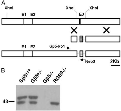
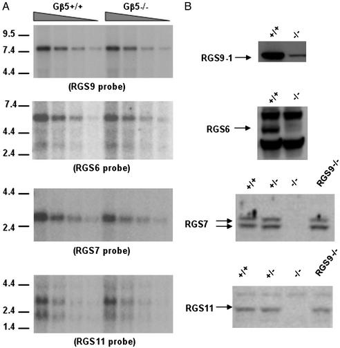
RGS (regulator of G protein signaling) proteins containing the G protein gamma-like (GGL) domain (RGS6, RGS7, RGS9, and RGS11) interact with the fifth member of the G protein beta-subunit family, Gbeta5. This interaction is necessary for the stability of both the RGS protein and for Gbeta5. Consistent with this notion, we have found that elevation of RGS9-1 mRNA levels by transgene expression does not increase RGS9-1 protein level in the retina, suggesting that Gbeta5 levels may be limiting. To examine further the interactions of Gbeta5 and the GGL domain-containing RGS proteins, we inactivated the Gbeta5 gene. We found that the levels of GGL domain-containing RGS proteins in retinas and in striatum are eliminated or reduced drastically, whereas the levels of Ggamma2 and RGS4 proteins remain normal in the absence of Gbeta5. The homozygous Gbeta5 knockout (Gbeta5-/-) mice derived from heterozygous knockout mating are runty and exhibit a high preweaning mortality rate. We concluded that complex formation between GGL domain-containing RGS proteins and the Gbeta5 protein is necessary to maintain their mutual stability in vivo. Furthermore, in the absence of Gbeta5 and all four RGS proteins that form protein complexes with Gbeta5, the animals that survive into adulthood are viable and have no gross defects in brain or retinal morphology.


Figures

Fig. 1.
Fig. 1.
The presence of Gβ5-S in the retina and striatum of RGS9/ mice. Retinal extracts (20 μg) and striatal extracts (CPu, 100 μg) from wild-type and RGS9/ mice (indicated, Top) were resolved by 12% SDS/PAGE, and the presence of Gβ5 and RGS9 spliced forms were visualized by Western blots by using generic RGS9 antibody (R4432) and Gβ5 antibody (CT-215). Molecular mass markers (top to bottom: 124, 81, 50, and 42 kDa) are shown on the left. The splice variants of RGS9 and Gβ5 are indicated by arrows (top to bottom, RGS9-2, RGS9-1, Gβ5-L, and Gβ5-S).
Fig. 2.
Fig. 2.
The generation of Gβ5/ mice. (A) Gβ5 gene-inactivation scheme. (Top) The first half of the mouse Gβ5 gene. (Middle) Targeting construct replacing a 2.7-kb XhoI fragment with a Neo marker (hatched block). (Bottom) The targeted locus can be identified by the presence of a 2.1-kb PCR fragment when amplified by using Neo3 and Gβ5-ko1 primers. (B) Western blotting of 20 μg of retinal extracts by using antibody CT-215 to show the presence of Gβ5 proteins in various genetic backgrounds. The long-spliced form, Gβ5-L, is absent in RGS9/ mouse retina, whereas both long and short forms of Gβ5 are absent in Gβ5/ mice.
Fig. 6.
Fig. 6.
RGS9-1 protein levels in TG9F transgenic mice. The pRho4.2-RGS9-1 transgenic construct shown in A was injected into mouse embryos to produce TG9F2 and TG9F10 mice that express full-length RGS9-1-coding region in the photoreceptors. The retinal levels of RGS9-1 transcripts were measured by dot blot as shown in B. The amounts of total retinal RNAs blotted on the filter (top to bottom) were 1, 0.5, 0.25, and 0.12 μg, respectively. The RGS9-1 message levels (Left) in TG9F2 and TG9F10 retinas were ≈2- and 8-fold more than that of the wild-type control. β-Actin was used as a control for loading (Right). (C) The RGS9-1 protein levels were determined by immunoblots of equal amount of retinal extracts. (D) Western blot analyses showing the amounts of RGS9-1 (Left) and Gβ5 spliced forms (Right) in retinas of indicated mouse lines. To determine whether the transgenic RGS9-1 mRNA could be translated, TG9F2 and TG9F10 mice were bred into the RGS9 knockout background to produce TG9F2/ and TG9F10/ mouse lines, respectively. The RGS9-1 protein level in TG9F2/ retina was ≈20% of the wild-type level, whereas in TG9F10/ it was 100%. A corresponding restoration of Gβ5-L in both TG9F2/ and TG9F10/ mouse retinas was evident. Molecular markers (in kilodaltons) are indicated on the right.
Fig. 3.
Fig. 3.
The growth of Gβ5/ mice in the first month of age. The mean body weights of the Gβ5/ (▵, n = 12) and Gβ5+/+ (▪, n = 4) mice generated in different litters were monitored daily after birth during their first month of growth. All pups were weaned on postnatal day 21 (indicated by an arrow). The smaller body size of the Gβ5/ pups is evident. In addition, a period of no weight gain, from postnatal day 15 to 20 (indicated by a bar) in the Gβ5/ pups was noticed. Error bars represent SD.
Fig. 4.
Fig. 4.
Instability of GGL domain-containing RGS proteins in retinas of Gβ5/ mice. (A) Retinal total RNA (3, 1.5, 0.75, and 0.38 μg from left to right) isolated from Gβ5/ and Gβ5+/+ mice was hybridized with various radioactive probes specific for RGS9, RGS6, RGS7, and RGS11 by Northern blotting (see Materials and Methods). The mRNA levels of the four RGS proteins were comparable between Gβ5/ and wild-type retinas. Molecular mass markers (in kb) are indicated at right. (B) Western blotting of retinal protein extracts derived from different genetic backgrounds by using antibodies specific for individual RGS proteins demonstrating the loss of RGS6, RGS7, and RGS11 and the reduction of RGS9-1 signals (indicated by arrows) in the Gβ5/ retina. Approximately 20 μg of retinal extract was analyzed for RGS7, RGS11, and RGS9-1; 40 μg was analyzed for RGS6 expression.
Fig. 5.
Fig. 5.
The absence of RGS9-2 and RGS7 but not RGS4 and Gγ2 in the striatum of Gβ5/ mice. (A) Northern blotting of total RNA (3, 1.5, 0.75, and 0.38 μg, left to right) isolated from Gβ5/ and Gβ5+/+ striatum was hybridized with radioactive RGS9 (Upper) and RGS7 (Lower) specific probes and visualized by autoradiography. The level of the 2.6-kb RGS9-2 transcript and the 2.7-kb RGS7 transcript was similar in Gβ5/ and in wild-type striatum. Molecular mass markers (in kilobases) are indicated at left. (B and C) Western blotting of 100 μg of striatal extracts by using antibodies for RGS9 (R4432), RGS7 (2923AP), RGS4 (SC-6204), and Gγ2 (SC-374), showing the specific loss of the 82-kDa RGS9-2 and the 55-kDa RGS7 signals in Gβ5/ mice. The levels of a non-GGL domain RGS protein, RGS4, and a regular γ-subunit, Gγ2, are not affected by the absence of Gβ5. Molecular mass markers (in kilodaltons) are indicated on the left.

References

    1. Dohlman, H. G., Song, J., Ma, D., Courchesne, W. E. & Thorner, J. (1996) Mol. Cell. Biol. 16, 5194–5209. - PMC - PubMed
    1. Koelle, M. R. & Horvitz, H. R. (1996) Cell 84, 115–125. - PubMed
    1. Berman, D. M., Wilkie, T. M. & Gilman, A. G. (1996) Cell 86, 445–452. - PubMed
    1. Ross, E. M. & Wilkie, T. M. (2000) Annu. Rev. Biochem. 69, 795–827. - PubMed
    1. Zheng, B., De Vries, L. & Gist Farquhar, M. (1999) Trends Biochem. Sci. 24, 411–414. - PubMed

Publication types

LinkOut - more resources