Review
. 2003 Apr;202(4):327-42.
doi: 10.1046/j.1469-7580.2003.00168.x.

Septation and separation within the outflow tract of the developing heart

Affiliations
Review

Septation and separation within the outflow tract of the developing heart

Sandra Webb et al. J Anat. 2003 Apr.

Abstract

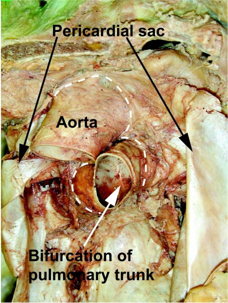
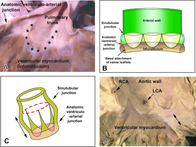
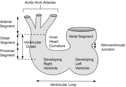
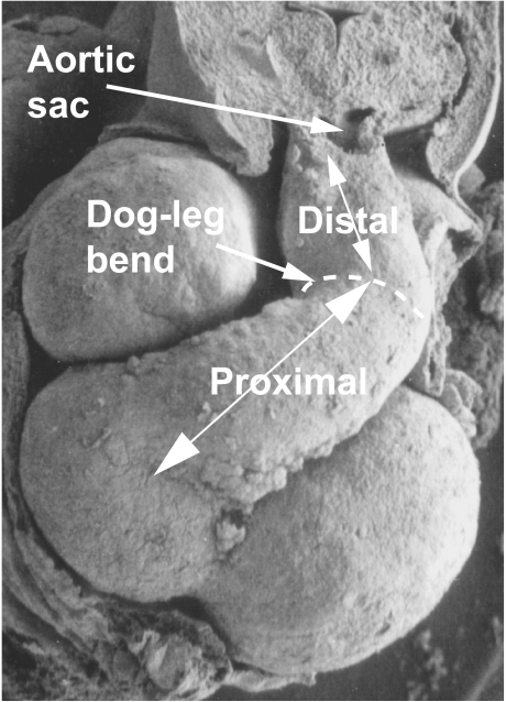
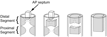
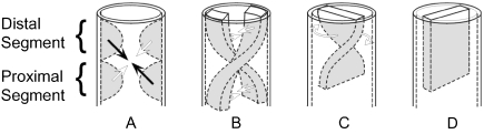
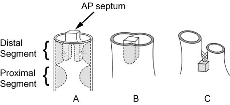
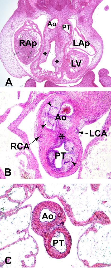
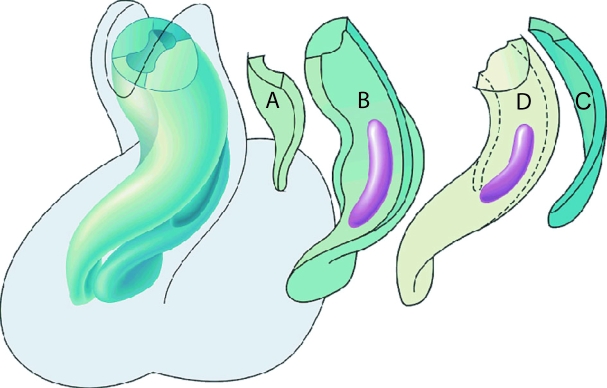
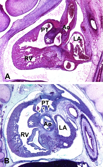
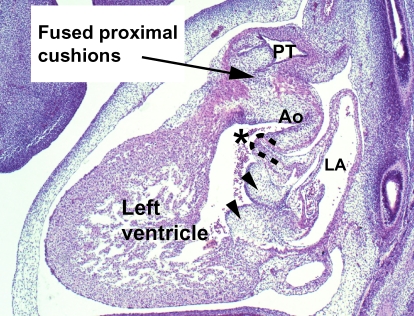
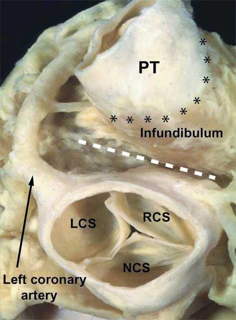
The developmental anatomy of the ventricular outlets and intrapericardial arterial trunks is a source of considerable confusion. First, major problems exist because of the multiple names and definitions used to describe this region of the heart as it develops. Second, there is no agreement on the boundaries of the described components, nor on the number of ridges or cushions to be found dividing the outflow tract, and the pattern of their fusion. Evidence is also lacking concerning the role of the fused cushions relative to that of the so-called aortopulmonary septum in separating the intrapericardial components of the great arterial trunks. In this review, we discuss the existing problems, as we see them, in the context of developmental and postnatal morphology. We concentrate, in particular, on the changes in the nature of the wall of the outflow tract, which is initially myocardial throughout its length. Key features that, thus far, do not seem to have received appropriate attention are the origin, and mode of separation, of the intrapericardial portions of the arterial trunks, and the formation of the walls of the aortic and pulmonary valvar sinuses. Also as yet undetermined is the formation of the free-standing muscular subpulmonary infundibulum, the mechanism of its separation from the aortic valvar sinuses, and its differentiation, if any, from the muscular ventricular outlet septum.


Figures

Fig. 1
Fig. 1
Heart removed from a human cadaver to show the extent of the aorta and the pulmonary trunk within the pericardial cavity. The dashed lines show the distal attachments of the fibrous pericardium.
Fig. 2
Fig. 2
(A) Pulmonary valve from an infant human heart. The valvar leaflets themselves have been removed to show the semilunar mode of their attachments (asterisks). Note the level of the anatomic ventriculoarterial junction between the arterial walls of the pulmonary trunk and the muscular right ventricular infundibulum. (B) Schematic drawing showing the mode of attachment of the valvar leaflets as shown in A, and their relationship to the sinutubular and ventriculoarterial junctions. Note that the bases of the valve leaflets overlap the ventricular myocardium. (C) Relationships of the valvar junctions and the valvar leaflets in three dimensions. Importantly, it demonstrates that the arterial valves have length (double headed arrows). (D) Opened aortic valve from an adult human heart subsequent to removal of the leaflets. As with the pulmonary valve, the leaflets are attached in semilunar fashion, but there is fibrous continuity between the non-coronary and left coronary leaflets of the aortic valve and the aortic leaflet of the mitral valve (double headed arrow). LCA, left coronary artery; RCA, right coronary artery.
Fig. 3
Fig. 3
This diagram of the developing embryonic heart illustrates our suggested terminology. Note that we define proximal and distal segments of the outflow tract. In the definitive situation, as shown in Fig. 4, the boundary between these components is marked by the characteristic bend ‘dog-leg’ bend. The bend has been ignored for the purposes of this drawing. The boundary between the distal outflow segment and the arterial segment, the aortic sac, is at the level marked by the reflections of the pericardial cavity (see also Fig. 1).
Fig. 4
Fig. 4
This scanning electron micrograph of a human embryo, at Carnegie stage 15 (approximately 34 days of gestation), shows a ventral view of the heart. The distal and proximal segments of the unseptated outflow tract are separated by a characteristic dog-leg bend. The proximal segment lies across the atrioventricular junction.
Fig. 5
Fig. 5
(A) Section through the outflow tract of a human embryo at Carnegie stage 14 (approximately 36 days of gestation) sectioned in the sagittal plane. It shows how the distal outflow tract with its myocardial wall (m) extends to the edge of the pericardial cavity, where it becomes continuous with the aortic sac. The aorto-pulmonary septum is seen as a wedge of tissue in the posterior wall of the sac (asterisk), separating the origins of the fourth and sixth aortic arches (4,6), the latter seen giving rise to one pulmonary artery. The outflow tract is being septated by the distal cushions. (B) Section from a human embryo at Carnegie stage 16 (approximately 37 days of gestation), which has been sectioned in the transverse plane. The distal cushions (asterisk) are shown separating into the intrapericardial parts of the aorta and the pulmonary trunk. Note the incipient arterialization of the walls of these trunks. The proximal outflow tract has retained its myocardial phenotype, proximal to the arrowheads.
Fig. 6
Fig. 6
Model of development whereby three components, namely the distal cushions, the proximal cushions and the aortopulmonary septum (AP septum), contribute to the septation of the outflow tract. Only those components that give rise to the septal structures are shown, the valvar leaflets having been excluded for simplicity. The panels show the presumed steps in septation.
Fig. 7
Fig. 7
Models for spiral septation of the outflow tract. The arrows in panel A indicate the fusion of opposite distal and proximal cushions to form spiralling longitudinal ridges, as shown in panel B. Others, however, argue that the spiralling ridges exist through the length of the outflow tract from the outset (see text for discussion). Be that as it may, having fused to form a spiralling septum, it is argued that ‘detorsion’ (panel C) is required to produce the definitive septum (panel D).
Fig. 8
Fig. 8
Diagram illustrating the concept of separation of the outflow tracts primarily by the aortopulmonary septum. Panel A shows the distal cushions fusing with the aortopulmonary septum to form a common septal structure (panel B). By the stage shown in C, the proximal cushions remain only as the posterior wall of the subpulmonary infundibulum (grey cube), with the aortopulmonary septum having reduced to a remnant of tissue external to the heart (cross-hatched).
Fig. 9
Fig. 9
(A) Frontal section through a human embryo at Carnegie stage 14 (approximately 35 days of gestation). The dorsal outflow tract has been separated into the interpericardial parts of the aorta (Ao) and the pulmonary trunk (PT). The proximal cushions (asterisks) have yet to fuse, but the dense mesenchymal tissue that originates from the neural crest is penetrating both cushions. RAp, LAp: right and left atrial appendages; LV, left ventricle. (B) Transverse section through a human embryo at Carnegie stage 22 (approximately 54 days of gestation) shows the stage subsequent to the septation of the distal outflow tract. Now the distal parts of the proximal cushions, together with the intercalated cushions, are cavitating (arrowheads) to form the valvar leaflets of the aorta (Ao) and the pulmonary trunk (PT), along with the walls of their supporting sinuses. Note the different stages of arterialization of the different sinusal walls, and note also that the sinuses remain enclosed within the myocardial cuff, through which the left (LCA) and right (RCA) coronary arteries are penetrating to enter the valvar sinuses. The dark staining fibrous tissue (asterisk) marks the initial site of fusion of the distal parts of the proximal cushions. (C) A more cranial slide from the same embryo, which shows that, by this stage, the intrapericardial parts of the arterial trunks are separate structures, with a bar of fibro-adipose tissue now occupying the former site of the distal cushions.
Fig. 10
Fig. 10
Reconstruction made from a human heart of 5 weeks gestation (Carnegie stage 15), viewed from the ventral aspect, soon after immigration of neural crest cells has begun. The left-hand panel shows the overall arrangement, with the arrangement of the individual cushions shown to the right. (A,C) Intercalated ridges or cushions, which occupy the area of the dog-leg bend; (B) septal outflow ridge; (D) parietal outflow ridge. Localized accumulations of neural crest-derived mesenchyme form ‘prongs’, shown in purple. Note that they are located within the distal parts of the cushions of the proximal outflow tract, being positioned just proximal to the dog-leg bend.
Fig. 11
Fig. 11
(A) Sagittal section from a human embryo at Carnegie stage 20 (approximately 50 days of gestation). The proximal cushions of the outflow tract have fused to form an embryonic outlet septum within the right ventricle (asterisk). The interventricular foramen, which links the right ventricle to the subaortic outflow, is seen as a channel positioned caudal to this outlet septum. The distal outflow segment has separated into the aortic and pulmonary trunks, each having an arterial phenotype. (B) Sagittal section from a human embryo of 11 weeks gestation. An extracardiac bar of tissue is now seen between the walls of the pulmonary trunk and the sinuses of the aorta (arrowheads). The proximal cushions have now myocardialized to form the subpulmonary infundibulum (asterisk). Abbreviations: Ao, aorta; LA, left atrium; PT, pulmonary trunk; RV, right ventricle.
Fig. 12
Fig. 12
Section from a human embryo at Carnegie stage 17 (approximately 44 days of gestation), sectioned in the sagittal plane. It shows that, although the aortic valve is being sequestered within the left ventricle by fusion of the proximal parts of the outflow cushions to the crest of the muscular ventricular septum, the musculature of the inner heart curvature (dashed line) still separates the developing leaflets of the aortic and mitral valves. The subaortic outlet is marked by an asterisk. The arrowheads indicate the atrioventricular endocardial cushions. AO, aorta; PT, pulmonary trunk; LA, left atrium.
Fig. 13
Fig. 13
This dissection of an adult human heart shows the relationship of the definitive ventricular outflow tracts. The arterial trunks have been removed, and the base of the heart is viewed from above. The anatomic pulmonary–ventricular junction, between the arterial wall of the pulmonary trunk (PT) and the muscular right ventricular infundibulum, is shown by the line of asterisks. Note the deep tissue plane (white dashed line) that separates the free-standing subpulmonary infundibulum of the right ventricle from the wall of the right coronary sinus of the aorta. Note also the left coronary artery. LCS, RCS, NCS – left, right, and non-coronary sinuses of the aorta, respectively.

Similar articles

Cited by

References

    1. Anderson RH. The anatomy of arteial valvar stenosis. Int. J. Cardiol. 1990;26:355–359. 10.1046/j.1469-7580.2003.00168.x. - DOI
    1. Anderson RH, Wilkinson JL, Arnold R, Lubkiewicz K. Morphogenesis of Bulboventricular malformations I: Consideration of embryogenesis in the normal heart. Br. Heart J. 1974a;36:2–42. 10.1046/j.1469-7580.2003.00168.x. - DOI - PMC - PubMed
    1. Anderson RH, Wilkinson JL, Arnold R, Becker AE, Lubkiewicz K. Morphogenesis of bulboventricular malformations II: observations on malformed hearts. Br. Heart J. 1974b;36:948–970. 10.1046/j.1469-7580.2003.00168.x. - DOI - PMC - PubMed
    1. Arguello C, De La Cruz MV, Sanchez C. Ultrastructural and experimental evidence of myocardial cell differentiation into connective tissue cells in embryonic chick heart. J. Mol. Cell Cardiol. 1978;10:307–315. 10.1046/j.1469-7580.2003.00168.x. - DOI - PubMed
    1. Arráez-Aybar La González-Lorrio F, Marantos-Gamarra Dg Jiménez-Collado J. Cardiac developmental onomatology: the real heart of the matter. J. Anat. 2003 in press. - PubMed

MeSH terms

LinkOut - more resources