. 2003 Apr;202(4):387-96.
doi: 10.1046/j.1469-7580.2003.00169.x.

Early GABA(A) receptor clustering during the development of the rostral nucleus of the solitary tract

Affiliations

Early GABA(A) receptor clustering during the development of the rostral nucleus of the solitary tract

W L Heck et al. J Anat. 2003 Apr.

Abstract

While there is an abundance of gamma-aminobutyric acid (GABA) in the gustatory zone of the nucleus of the solitary tract of the perinatal rat, we know that GABAergic synapse formation is not complete until well after birth. Our recent results have shown that GABA(B) receptors are present at birth in the cells of the nucleus; however, they do not redistribute and cluster at synaptic sites until after PND10. The present study examined the time course of appearance and redistribution of GABA(A) receptors in the nucleus. GABA(A) receptors were also present at birth. However, in comparison to GABA(B) receptors, GABA(A) receptors underwent an earlier translocation to synaptic sites. Extrasynaptic label, for example, of GABA(A) receptors was non-existent compared to GABA(B) receptors at PND10 and well-defined clusters of GABA(A) receptors could be seen as early as PND1. We propose that while GABA(A), receptors may play an early neurotransmitter role at the synapse, GABA(B) receptors may play a non-transmitter neurotrophic role.


Figures
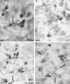
Fig. 1
Fig. 1
Light microscopic images of immunoreactivity for GABAA receptors in PND1 (A), PND 5 (B), PND 10 (C) and adult (D) rat rNST. Immunoreactivity is present in somata (asterisks) and extends into the dendritic processes (arrows) at all ages. Immunoreactivity is present right after birth in the rat in many cells, but the clusters (arrowheads) do not become common until PND5. On PND1, a few large neurones show the clustered pattern of immunoreactivity (one example is seen in the centre of this figure). The distinct clustering of immunoreactivity is present in each labelled cell by PND10 and remains in the adult.
Fig. 2
Fig. 2
Histogram of the mean density (darkness of label per unit area) of GABAA receptor immunoreactivity in rNST neurones during postnatal development. Somatic staining for GABAA receptors is highest at PND1, decreases to PND5 (P < 0.0001) and increases again to be maintained at adult levels by PND10 (P < 0.0001). This graph could be contrasted with Fig. 5 in Heck et al. (2001), the analogous graph for GABAB receptors in the rat rNST. The same pattern is seen; however, GABAB receptor development is delayed compared to GABAA. For GABAB receptors the decrease in overall immunoreactivity occurs between PND10 and PND15 and the increase to adult levels occurs after weaning (PND20). GABAA receptors mature earlier than GABAB receptors. Vertical bars indicate the standard errors of the mean. Horizontal arrows with stars indicate significant differences between the groups as determined with Fisher post hoc PLSD tests.
Fig. 3
Fig. 3
Electron micrographs of GABAA receptor labelling in the adult (A,B,C) and PND10 (D,E) rat rNST. The ultrastructure is well preserved in this tissue despite the fact that it was fixed to preserve immunogenicity. Background label is very scarce and labelled synapses (arrows) are easy to detect. Label is found either in the cleft or in association with the post-synaptic density. The targets of synapses shown are a large dendrite (A) and two small dendrites (B,C). Even at PND10, the ultrastructure is well preserved (D). Labelled synapses appear very mature, although the density of vesicles is somewhat low and the post-synaptic targets are quite large (and are probably quite proximal to the cell body, see text). Panel E shows a clathrin-coated vesicle pinching off near the synapse. Such structures have been noted before in association with post-synaptic membranes of developing GABAergic terminals (see Brown et al. 2000).

References

    1. Belhage B, Hansen GH, Elster L, Schousboe A. Effects of GABA on synaptogenesis and synaptic function. Perspectives Dev. Neurobiol. 1998;5:235–246. - PubMed
    1. Bevan S, Steinbach JH. The distribution of α-bungarotoxin binding sites on mammalian skeletal muscle developing in vivo. J. Physiol. 1977;267:195–213. - PMC - PubMed
    1. Bradley RM, King MS, Wang L, Shu X. Neurotransmitter and neuromodulator activity in the gustatory zone of the nucleus tractus solitarius. Chem. Senses. 1996;21:377–385. - PubMed
    1. Brandon NJ, Bedford FK, Connolly CN, Couve A, Kittler JT, Hanley JG, et al. Synaptic targeting and regulation of GABA (A) receptors. Biochem. Soc. Transactions. 1999;27:527–530. - PubMed
    1. Brown M, Renehan WE, Schweitzer L. Changes in GABA-immunoreactivity during development of the rostral subdivision of the nucleus of the solitary tract (rNST) Neuroscience. 2000;100:849–859. - PubMed

Publication types

Substances

LinkOut - more resources