

Transthoracic Doppler echocardiography - noninvasive diagnostic window for coronary flow reserve assessment
- PMID: 12740038
- PMCID: PMC155634
- DOI: 10.1186/1476-7120-1-4
Transthoracic Doppler echocardiography - noninvasive diagnostic window for coronary flow reserve assessment
Abstract
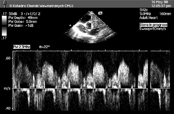
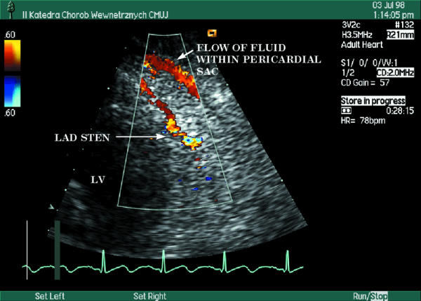
This review focuses on transthoracic Doppler echocardiography as noninvasive method used to assess coronary flow reserve (CFR) in a wide spectrum of clinical settings. Transthoracic Doppler echocardiography is rapidly gaining appreciation as popular tool to measure CFR both in stenosed and normal epicardial coronary arteries (predominantly in left anterior descending coronary artery). Post-stenotic CFR measurement is helpful in: functional assessment of moderate stenosis, detection of significant or critical stenosis, monitoring of restenosis after revascularization. In the absence of stenosis in the epicardial coronary artery, decreased CFR enable to detect impaired microvascular vasodilatation in: reperfused myocardial infarct, arterial hypertension with or without left ventricular hypertrophy, diabetes mellitus, hypercholesterolemia, syndrome X, hypertrophic cardiomyopathy. In these diseases, noninvasive transthoracic Doppler echocardiography allows for serial CFR evaluations to explore the effect of various pharmacological therapies.
Figures

Similar articles
-
Assessment of flow velocity reserve by transthoracic Doppler echocardiography and venous adenosine infusion before and after left anterior descending coronary artery stenting.J Am Coll Cardiol. 2001 Jul;38(1):155-62. doi: 10.1016/s0735-1097(01)01333-x. J Am Coll Cardiol. 2001. PMID: 11451266
-
Functional assessment of the collateral-dependent circulation in chronic total coronary occlusion using transthoracic Doppler ultrasound and venous adenosine infusion.Am J Cardiol. 2006 Jul 15;98(2):197-203. doi: 10.1016/j.amjcard.2006.01.075. Epub 2006 May 24. Am J Cardiol. 2006. PMID: 16828592
-
Noninvasive evaluation of flow reserve in the left anterior descending coronary artery in patients with cardiac syndrome X.Am J Cardiol. 2007 May 15;99(10):1378-83. doi: 10.1016/j.amjcard.2006.12.070. Epub 2007 Mar 28. Am J Cardiol. 2007. PMID: 17493464
-
Clinical application of transthoracic Doppler echocardiography to assess coronary flow reserve.Przegl Lek. 2002;59(8):629-31. Przegl Lek. 2002. PMID: 12638336 Review.
-
[Non-invasive assessment of coronary flow reserve with transthoracic echocardiography: physiopathology, methodology and clinical value].Ital Heart J Suppl. 2003 Mar;4(3):179-88. Ital Heart J Suppl. 2003. PMID: 12784752 Review. Italian.
Cited by
-
Prognostic Value of Coronary Flow Reserve Obtained on Dobutamine Stress Echocardiography and its Correlation with Target Heart Rate.Arq Bras Cardiol. 2017 May;108(5):417-426. doi: 10.5935/abc.20170041. Epub 2017 Apr 20. Arq Bras Cardiol. 2017. PMID: 28444062 Free PMC article.
-
Association between serum total antioxidant status and coronary microvascular function in idiopathic dilated cardiomyopathy.Herz. 2015 May;40(3):487-94. doi: 10.1007/s00059-013-4021-x. Epub 2014 Mar 9. Herz. 2015. PMID: 24609797
-
Reduced coronary flow reserve in Anderson-Fabry disease measured by transthoracic Doppler echocardiography.Cardiovasc Ultrasound. 2005 Apr 27;3:11. doi: 10.1186/1476-7120-3-11. Cardiovasc Ultrasound. 2005. PMID: 15857518 Free PMC article.
-
Coronary flow reserve in stress-echo lab. From pathophysiologic toy to diagnostic tool.Cardiovasc Ultrasound. 2005 Mar 25;3:8. doi: 10.1186/1476-7120-3-8. Cardiovasc Ultrasound. 2005. PMID: 15792499 Free PMC article. Review.
-
The non-invasive documentation of coronary microcirculation impairment: role of transthoracic echocardiography.Cardiovasc Ultrasound. 2005 Aug 4;3:18. doi: 10.1186/1476-7120-3-18. Cardiovasc Ultrasound. 2005. PMID: 16080792 Free PMC article. Review.
References
-
- Dimitrow PP. Coronary flow reserve-measurement and application: focus on transthoracic Doppler echocardiography. Boston/Dordrecht/London: Kluwer Academic Publishers. 2002.
-
- Picano E. Stress echocardiography: a historical perspective. Am J Med. 2003;114:126–130. - PubMed
-
- Radvan J, Marwick TH, Williams MJ, Camici PG. Evaluation of the extent and timing of the coronary hyperemic response to dipyridamole: a study with transesophageal echocardiography and positron emission tomography with oxygen 15 water. J Am Soc Echocardiogr. 1995;8:864–873. - PubMed
-
- Reis SE, Holubkov R, Lee JS, Sharaf B, Reichek N, Rogers WJ, Walsh EG, Fuisz AR, Kerensky R, Detre KM, Sopko G, Pepine CJ. Coronary flow velocity response to adenosine characterizes coronary microvascular function in women with chest pain and no obstructive coronary disease. Results from the pilot phase of the Women's Ischemia Syndrome Evaluation (WISE) study. J Am Coll Cardiol. 1999;33:1469–1475. - PubMed
-
- Saraste M, Koskenvuo J, Knuuti J, Toikka J, Laine H, Niemi P, Sakuma H, Hartiala J. Coronary flow reserve: measurement with transthoracic Doppler echocardiography is reproducible and comparable with positron emission tomography. Clin Physiol. 2001;21:114–122. - PubMed
Publication types
MeSH terms
LinkOut - more resources
Full Text Sources
