. 2003 Jul 1;550(Pt 1):241-53.
doi: 10.1113/jphysiol.2003.038356. Epub 2003 May 9.

Evolution of learning in three aplysiid species: differences in heterosynaptic plasticity contrast with conservation in serotonergic pathways

Affiliations

Evolution of learning in three aplysiid species: differences in heterosynaptic plasticity contrast with conservation in serotonergic pathways

Stéphane Marinesco et al. J Physiol. .

Abstract

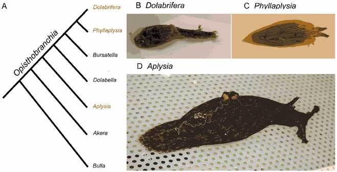
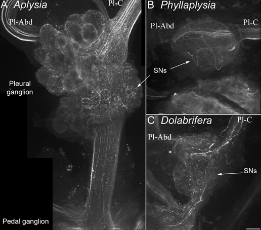
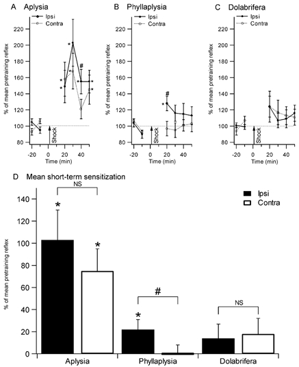
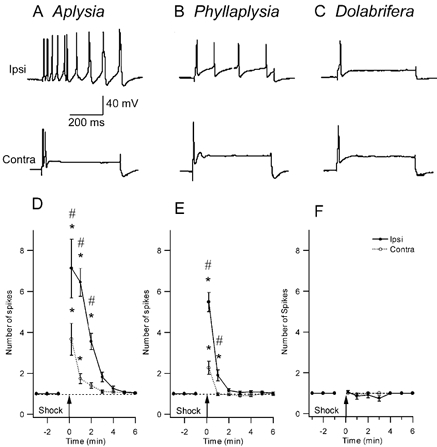
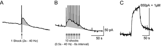
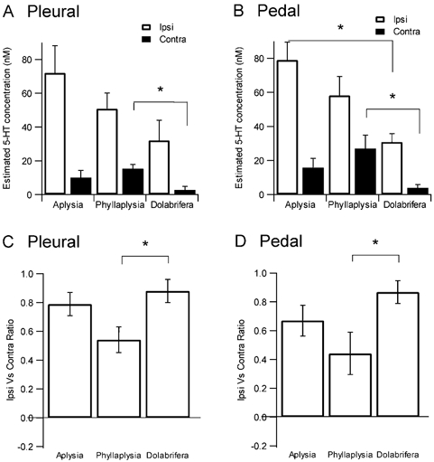
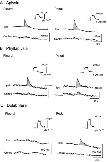
We investigated the neurobiological basis of variation in sensitization between three aplysiid species: Aplysia californica, Phyllaplysia taylori and Dolabrifera dolabrifera. We tested two different forms of sensitization induced by a noxious tail shock: local sensitization, expressed near the site of shock, and general sensitization, tested at remote sites. Aplysia showed both local and general sensitization, whereas Phyllaplysia demonstrated only local sensitization, and Dolabrifera lacked both forms of learning. We then investigated a neurobiological correlate of sensitization, heterosynaptic modulation of sensory neuron excitability by tail-nerve stimulation. We found (1) an increase in sensory neuron (SN) excitability after both ipsilateral and contralateral nerve stimulation in Aplysia, (2) a smaller and shorter-lasting increase in Phyllaplysia, and (3) no effect in Dolabrifera. Because sensitization in Aplysia is strongly correlated with serotonergic (5-HT) neuromodulation, we hypothesized that the observed interspecific variation in sensitization and SN neuromodulation might be correlated with variation in the anatomy and/or functional response of the serotonergic system. However, using immunohistochemistry, we found that all three species showed a similar pattern of 5-HT innervation. Furthermore, they also showed comparable 5-HT release evoked by tail-nerve shock, as measured with chronoamperometry. These observations indicate that interspecific variation in learning is correlated with differences in SN heterosynaptic plasticity within a background of evolutionary conservation in the 5-HT neuromodulatory pathway. We thus hypothesize that evolutionary changes in learning phenotype do not involve modifications of the 5-HT pathway per se, but rather, changes in the response of SNs to the activation of this or other neuromodulatory pathways upon noxious stimulation.


Figures

Figure 1
Figure 1. Evolution of opistobranch species
A, cladogram of the evolution of opistobranch species (adapted from a recent cladistic analysis of mDNA sequence and conventional character traits; Medina & Walsh, 2000). All species except Bulla are in the family Aplysiidae. B-D, pictures of the different species used in the present study: Dolabrifera (B), Phyllaplysia (C) and Aplysia (D). Wild-caught adult Aplysia individuals are usually much larger (about 250 g body weight, 15 cm long) than their Dolabrifera and Phyllaplysia counterparts (2–3 g body weight, 3 cm long).
Figure 4
Figure 4. Immunohistochemical labelling of serotonin-immunoreactive fibres in the vicinity of tail sensory neurons
Confocal photomicrographs of clusters of tail SNs in the pleural ganglion, stained with an anti-5-HT antibody were taken from Aplysia (A), Phyllaplysia (B) and Dolabrifera (C, n = 2). The orientation is the same for all species: pleural ganglion (top), pedal ganglion (bottom), pleural-abdominal nerve (Pl-Abd, left) and pleural-cerebral nerve (Pl-C, right). The three species show a similar pattern of 5-HT innervation: numerous 5-HT fibres are present in the cerebral-pleural nerve, they branch over the cluster of tail SNs (arrows) and/or travel to the abdominal ganglion through the pleural-abdominal nerve. Some 5-HT processes also travel towards the pedal ganglion through the pleural-pedal nerve. No 5-HT-immunoreactive cell bodies were evidenced in the pleural ganglion. Scale bar = 100 μm.
Figure 2
Figure 2. Sensitization of the tail withdrawal reflex in the three species
Reflex duration (% of mean pretraining reflex) after training is shown in the top panels (A, B, and C). Tail withdrawal was tested every 10 min before and after tail shock in Aplysia (n = 11, A), Phyllaplysia (n = 12, B) and Dolabrifera (n = 11, C). Combined post-shock data are displayed in D. Aplysia showed significant sensitization both ipsilateral and contralateral to the shock. Phyllaplysia showed only local (ipsilateral) sensitization. Dolabrifera showed a transient, reduced form of sensitization on both sides. Analysis of short-term sensitization (mean between 20 and 30 min tests) confirmed the presence of both local and general sensitization in Aplysia, and local sensitization only in Phyllaplysia. However, the small enhancement in tail withdrawal in Dolabrifera was not significant (D). *Significant difference between pre- and post-test values (P < 0.05); # significant difference between ipsilateral and contralateral sides (P < 0.05).
Figure 3
Figure 3. Comparison of the effect of nerve shock on excitability of tail sensory neurons in the three study species
A-C, representative examples of intracellular recordings from homologous SNs in three species. The sensory neuron was injected with a 500 ms intracellular pulse of depolarizing current, 1 min after electrical stimulation of a peripheral tail nerve (top trace: ipsilateral tail-nerve shock; bottom trace: contralateral tail-nerve shock). Current amplitude was set prior to nerve stimulation to give a single action potential (traces not shown). D-F, SN excitability across time after nerve stimulation shows the change in the number of evoked spikes after nerve shock. D, Aplysia (n = 17) shows a significant increase in excitability both ipsi- and contralateral to the shock, that lasts about 3–5 min. E, in Phyllaplysia (n = 17), nerve shock increased excitability only in the ipsilateral SNs for 1 min after nerve stimulation. F, in Dolabrifera (n = 11), no excitability increase was observed after nerve shock. * Significant difference between pre- and post-test values (P < 0.05); # significant difference between ipsilateral and contralateral sides (P < 0.05).
Figure 5
Figure 5. 5-HT release evoked by one or repeated tail-nerve shocks in Aplysia
The tail nerve was stimulated using one single shock (A, 2 s train of 5 ms/30 V pulses at 40 Hz) or 10 shocks (B, 500 ms train of 5 ms/30 V pulses at 40 Hz) repeated at 6 s intervals. Time of shocks is shown by arrows. The vertical deflections in the 5-HT signal are caused by the stimulus artifact. C, calibration of the electrode used for the experiment in 5-HT 1 μM, using a flow injection chamber. 5-HT release is only slightly greater with repeated stimulation because of a severe depression in the 5-HT signals evoked by each successive stimulus.
Figure 7
Figure 7. Summary data and statistical analysis of 5-HT release evoked by tail-nerve stimulation
Top, maximal concentration of 5-HT release evoked by ipsilateral or contralateral tail-nerve stimulation in the pleural (A) and pedal (B) ganglia. Ipsilateral stimulation released a greater concentration of 5-HT into the extracellular space in Aplysia than in Phyllaplysia and Dolabrifera. However, 5-HT release evoked by contralateral stimulation was maximal in Phyllaplysia, intermediate in Aplysia and minimal in Dolabrifera. Bottom, a ratio of lateralization was computed as (Ipsi - Contra)/(Ipsi + Contra); 1.0 for complete lateralization to the ipsilateral side, 0.0 for equal release in ipsi- and contralateral sides, −1.0 for complete lateralization to the contralateral side. The lateralization ratio for both pleural (C) and pedal (D) ganglia was maximal for Dolabrifera, intermediate for Aplysia and minimal for Phyllaplysia. Histograms show mean ±s.e.m. *P ≤ 0.05 as assessed by a one-way ANOVA followed by a LSD post hoc test.
Figure 6
Figure 6. Examples of 5-HT release evoked by tail-nerve stimulation in Aplysia (A), Phyllaplysia (B) and Dolabrifera (C)
Chronoamperometric recordings obtained from carbon fibre electrodes implanted underneath tail SNs in the pleural ganglion (left) and in the ventral portion of the pedal ganglion proximal to the pleural-pedal nerve, in the region of tail SN-MN synapses (right). Serotonin release was evoked by ipsilateral (Ipsi) and contralateral (Contra) tail-nerve stimulation and 5-HT concentrations were estimated using the calibration value obtained in 5-HT 1 μM for each electrode (upper right of each recording). Consistent 5-HT release was found in all species, lasting 30–40 s after tail-nerve stimulation. However the maximal 5-HT concentration as well as the ratio between the amounts of 5-HT released by ipsilateral and contralateral stimulation varied between the three species. Increases in 5-HT oxidation currents have been underlined by shading, and stimulation artifacts have been removed.

Similar articles

Cited by

References

    1. Abrams TW, Castelluci VF, Camardo JS, Kandel ER, Lloyd PE. Two endogenous neuropeptides modulate the gill and siphon withdrawal reflex in Aplysia by presynaptic facilitation involving cAMP-dependent closure of a serotonin-sensitive potassium channel. Proc Natl Acad Sci U S A. 1984;81:7956–7960. - PMC - PubMed
    1. Baxter D, Byrne J. Serotonergic modulation of two potassium currents in the pleural sensory neurons of Aplysia. J Neurophysiol. 1989;62:665–679. - PubMed
    1. Belkin K, Abrams T. FMRFamide produces biphasic modulation of the LFS motor neurons in the neural circuit of the siphon withdrawal reflex of Aplysia by activating Na+ and K+ currents. J Neurosci. 1993;13:5139–5152. - PMC - PubMed
    1. Billy A, Walters E. Modulation of mechanosensory threshold in Aplysia by serotonin, small cardioactive peptide B (SCPB), FMRFamide, acetylcholine and dopamine. Neurosci Lett. 1989;105:200–204. - PubMed
    1. Bristol A, Fischer T, Carew T. Combined effects of intrinsic facilitation and modulatory inhibition of identified interneurons in the siphon withdrawal circuitry of Aplysia. J Neurosci. 2001;21:8990–9000. - PMC - PubMed

Publication types

LinkOut - more resources