. 2003 May 15;22(10):2453-62.
doi: 10.1093/emboj/cdg226.

Regulation of Smad signaling through a differential recruitment of coactivators and corepressors by ZEB proteins

Affiliations

Regulation of Smad signaling through a differential recruitment of coactivators and corepressors by ZEB proteins

Antonio A Postigo et al. EMBO J. .

Abstract

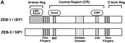
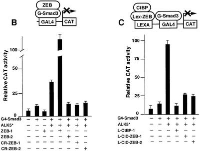
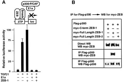
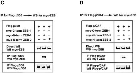
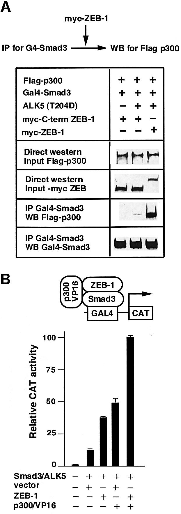
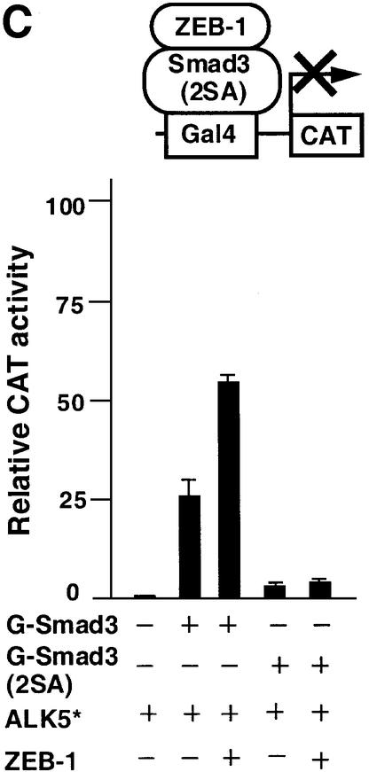
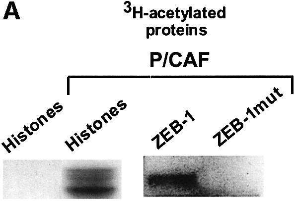
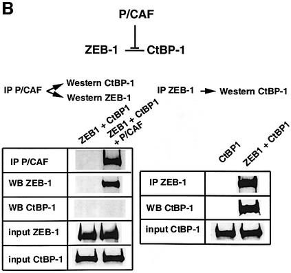
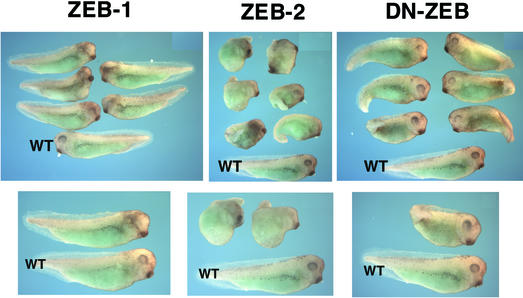
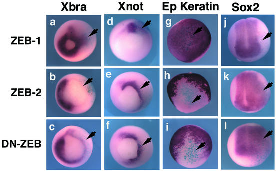
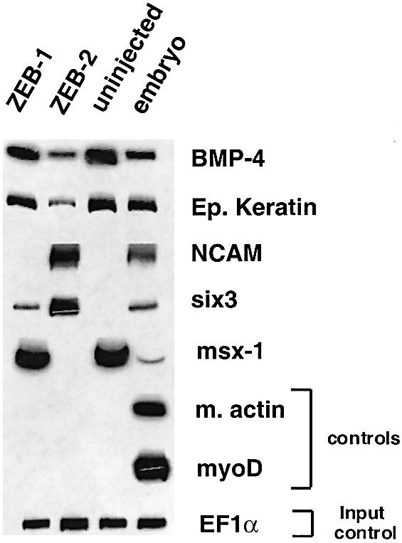
Balancing signals derived from the TGFbeta family is crucial for regulating cell proliferation and differentiation, and in establishing the embryonic axis during development. TGFbeta/BMP signaling leads to the activation and nuclear translocation of Smad proteins, which activate transcription of specific target genes by recruiting P/CAF and p300. The two members of the ZEB family of zinc finger factors (ZEB-1/deltaEF1 and ZEB-2/SIP1) regulate TGFbeta/BMP signaling in opposite ways: ZEB-1/deltaEF1 synergizes with Smad-mediated transcriptional activation, while ZEB-2/SIP1 represses it. Here we report that these antagonistic effects by the ZEB proteins arise from the differential recruitment of transcriptional coactivators (p300 and P/CAF) and corepressors (CtBP) to the Smads. Thus, while ZEB-1/deltaEF1 binds to p300 and promotes the formation of a p300-Smad transcriptional complex, ZEB-2/SIP1 acts as a repressor by recruiting CtBP. This model of regulation by ZEB proteins also functions in vivo, where they have opposing effects on the regulation of TGFbeta family-dependent genes during Xenopus development.


Figures

None
Fig. 1. (A) Schematic representation of the ZEB family (ZEB-1/δEF1 and ZEB-2/SIP1) of two-handed zinc finger factors. The central region (CR) in between both zinc finger clusters act as a repressor domain in part through the recruitment of the CtBP corepressor through a CtBP-interacting domain (CID). There is a region 3′ of the N-terminal zinc fingers that acts as a Smad-binding domain (SID). The N-terminal region of ZEB-1/δEF1 serves as a p300–p/CAF-binding domain (see Figure 2C). (B) The central repressor (CR) domain of both ZEB-1/δEF1 and ZEB-2/SIP1 inhibits the activity of Smad3 when brought directly to the promoter. 293T cells were transfected with 0.25 µg of a reporter construct (PG5-CAT) containing five Gal4 DNA-binding sites driving the CAT gene, 0.25 µg of Gal4–Smad3 and 0.1 µg of an expression vector for constitutively active ALK5 (T204D) (ALK5*) along with either 1 µg of expression vectors for either ZEB-1/δEF1 or ZEB-2/SIP1, 0.8 µg of either CR-ZEB-1 or CR-ZEB-2, or 0.7 µg of vector alone (CS2MT, not shown). (C) The CID of both ZEB proteins or CtBP-1 itself represses Smad3 transcriptional activation. 293T cells were cotransfected with 0.5 µg of a CAT reporter construct (PL6G5-CAT) containing both LexA and Gal4 DNA-binding sites along with 0.25 µg of Gal4–Smad3, 0.1 µg of an expression vector for constitutively active ALK5 (T204D) (ALK5*) and either 0.45 µg of LexA empty vector (PBXL3, not shown) or 0.5 µg of LexA-CtBP-1, LexA-CID-ZEB-1 or LexA-CID-ZEB-2 expression vectors. In all the above experiments, equal molar amounts of each empty vector were transfected as controls; 0.1 µg of SV40βgal was also cotransfected to control for transfection efficiency. Luciferase values were assessed as described in Material and methods.
None
Fig. 2. The N-terminal domain of ZEB-1/δEF1 interacts with coactivators p300 and P/CAF. (A) ZEB-1/δEF1 synergy with TGFβ requires the p300 coactivator. 3TP-luc reporter (0.3 µg) was transfected in Mv1Lu along with 0.3 µg of an expression vector for E1A 12S and either 0.48 µg of CS2MT empty vector (not shown) or 0.7 µg of CS2MT-ZEB-1 either in the absence or presence of 25 pM of TGFβ1. SV40βgal (0.1 µg) was cotransfected in all points to control for transfection efficiency. (B) Full-length ZEB-1/δEF1, but not ZEB-2/SIP1, interacts with p300. 293T cells were cotransfected with 10 µg of a Flag-tagged p300 expression vector and 20 µg of either myc-tagged ZEB-1/δEF1 or myc-tagged ZEB-2/SIP1 expression vectors. After 48 h, cells lysates were immunoprecipitated (IP) for Flag-p300 and the binding to ZEB-1/δEF1 (but not to ZEB-2/SIP1) determined by western blotting (WB) with an anti-myc 9E10 mAb. (C) The N-terminal domain of ZEB-1/δEF1 interacts with p300. 293T cells were cotransfected with 10 µg of Flag-tagged p300 expression vector and 5 µg of the indicated myc-tagged ZEB expression vectors. Cell lysates were immunoprecipitated for Flag-p300 and binding to the different regions of ZEB-1/δEF1 and ZEB-2/SIP1 determined by western blotting with an anti-myc 9E10 mAb. (D) P/CAF interacts with the N-terminal region of ZEB-1/δEF1 but not ZEB-2/SIP1. Experiments were performed similarly to those in (C) except that P/CAF was expressed instead of p300.
None
Fig. 3. ZEB-1/δEF1 promotes the assembly of a Smad3–p300 complex. (A) 293T cells were cotransfected with 5 µg of the indicated expression vectors and 0.8 µg of the constitutively active ALK5 (T204D). ZEB-1 contains the sequence of ZEB-1/δEF1 from the N-terminal region to the SID. After 48 h, cell lysates were immunoprecipitated (IP) for Smad3 and associated p300 was determined by western blotting (WB). The input (indicated as direct western) represents 15% of the lysate. (B) ZEB-1/δEF1 recruits p300 to Smad3. The PG5-CAT reporter containing five Gal4 sites fused to CAT (0.6 µg) was transfected in C33a cells along with the following expression vectors: 0.2 µg of Gal4–Smad3, 0.3 µg of constitutively active ALK5, either 0.48 µg of empty vector CS2MT or 0.7 µg of CS2MT-ZEB-1 and 0.25 µg of a p300–VP16 fusion protein. One-tenth of 1 µg of SV40βgal was cotransfected to control for transfection efficiency. CAT assays were performed as described in Materials and methods. (C) Formation of a p300–Smad3–ZEB-1/δEF1 complex requires the binding of p300 to Smad3. C33a cells were transfected with the same CAT reporter as in (B) along with either 0.2 µg of Gal4–Smad3 or Gal–Smad3 (2SA), 0.3 µg of constitutively active ALK5 (T204D) (ALK5*) and either 0.48 µg of empty vector CS2MT (not shown) or 0.7 µg of CS2MT-ZEB-1. One tenth of 1 µg of SV40βgal was cotransfected to control for transfection efficiency.
None
Fig. 4. P/CAF acetylates the lysine residues flanking the CID in ZEB-1/δEF1 and displaces CtBP. (A) Purified recombinant P/CAF was used in an in vitro assay to acetylate core histones or agarose beads coupled to either GST–CID-ZEB-1 (wild type) or GST–CID-ZEB-1 mutant (K741A, K774A, K775A). (B) Expression of P/CAF leads to displacement of CtBP-1 from ZEB-1/δEF1. The indicated tagged expression vectors (Flag-P/CAF, Gal4-ZEB-1/δEF1 and myc-CtBP) were transfected into 293T cells as in previous experiments and cell extracts were immunoprecipitated with antibodies to either Gal4-ZEB- 1/δEF1 or Flag-P/CAF. Note that the experiments were designed such that a similar amount of ZEB-1/δEF1 was immunoprecipitated directly (anti-Gal4-ZEB-1/δEF1) or co-immunoprecipitated by P/CAF (compare the left and right panels). No CtBP-1 was associated with ZEB-1/δEF1 when it was bound to P/CAF.
None
Fig. 5. Regulation of Xenopus development by ZEB-1/δEF1 and ZEB-2/SIP1 in vivo. Phenotype of Xenopus embryos injected at the two-cell stage with RNA for ZEB-1/δEF1, ZEB-2/SIP1 or DN-ZEB. Embryos were allowed to develop until tailbud (stage 29–30). Embryos injected with ZEB-2/SIP1 or DN-ZEB mRNA were significantly dorsalized. In contrast, ZEB-1/δEF1 showed no dorsalizing effect, and there was disruption of eye development in the injected side. In each panel, a wild-type embryo is shown at the bottom (indicated as WT).
None
Fig. 6. Regulation of activin/BMP-dependent genes and neural genes by ZEB-1/δEF1 and ZEB-2/SIP1. In situ hybridization for the indicated genes in embryos injected with ZEB-1/δEF1, ZEB-2/SIP1 or DN-ZEB RNAs along with mRNA encoding β-galactosidase (to mark the side of the injection) and allowed to develop to stage 11.5 (for expression of Xbrachyury, Xnot and epidermal keratin) or stage 13 (for Sox2). Whereas ZEB-1/δEF1 induced ectopic expression of Brachyury, ZEB-2/SIP1and DN-ZEB inhibited Brachyury expression. There was no effect on the control uninjected side (left side of each embryo).
None
Fig. 7. Differential effect of ZEB-1/δEF1 versus ZEB-2/SIP1 on gene expression. mRNA isolated from animal caps of Xenopus embryos (either uninjected or injected with mRNA for ZEB-1/δEF1 or ZEB-2/SIP1) was used for RT–PCR analysis of the indicated genes (see Materials and methods for details).

Similar articles

Cited by

References

    1. Bannister A.J. and Kouzarides,T. (1996) The CBP co-activator is a histone acetyltransferase. Nature, 384, 641–643. - PubMed
    1. Barlev N.A., Liu,L., Chehab,N.H., Mansfield,K., Harris,K.G., Halazonetis,T.D. and Berger,S.L. (2001) Acetylation of p53 activates transcription through recruitment of coactivators/histone acetyltransferases. Mol. Cell, 8, 1243–1254. - PubMed
    1. Candia A.F., Watabe,T., Hawley,S.H., Onichtchouk,D., Zhang,Y., Derynck,R., Niehrs,C. and Cho,K.W. (1997) Cellular interpretation of multiple TGF-β signals: intracellular antagonism between activin/BVg1 and BMP-2/4 signaling mediated by Smads. Development, 124, 4467–4480. - PubMed
    1. Chakravarti D., Ogryzko,V., Kao,H.Y., Nash,A., Chen,H., Nakatani,Y. and Evans,R,M. (1999) A viral mechanism for inhibition of p300 and PCAF acetyltransferase activity. Cell, 96, 393–403. - PubMed
    1. Chamberlain E.M. and Sanders,M.M. (1999) Identification of the novel player δEF1 in estrogen transcriptional cascades. Mol. Cell. Biol., 19, 3600–3606. - PMC - PubMed

Publication types

MeSH terms