. 2003 May 15;22(10):2495-504.
doi: 10.1093/emboj/cdg232.

Centromeric chromatin pliability and memory at a human neocentromere

Affiliations

Centromeric chromatin pliability and memory at a human neocentromere

Jeffrey M Craig et al. EMBO J. .

Abstract

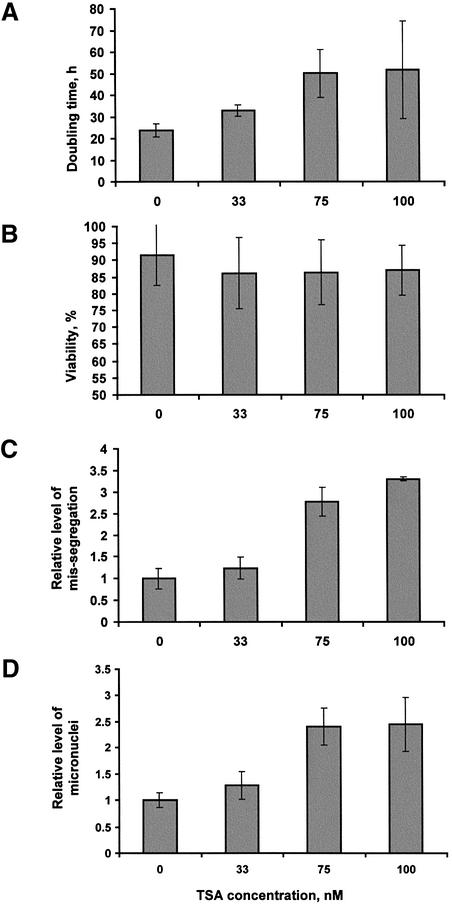
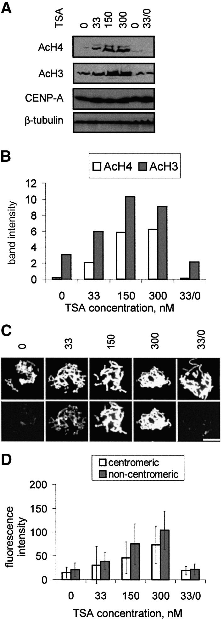
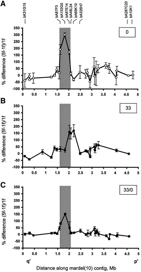
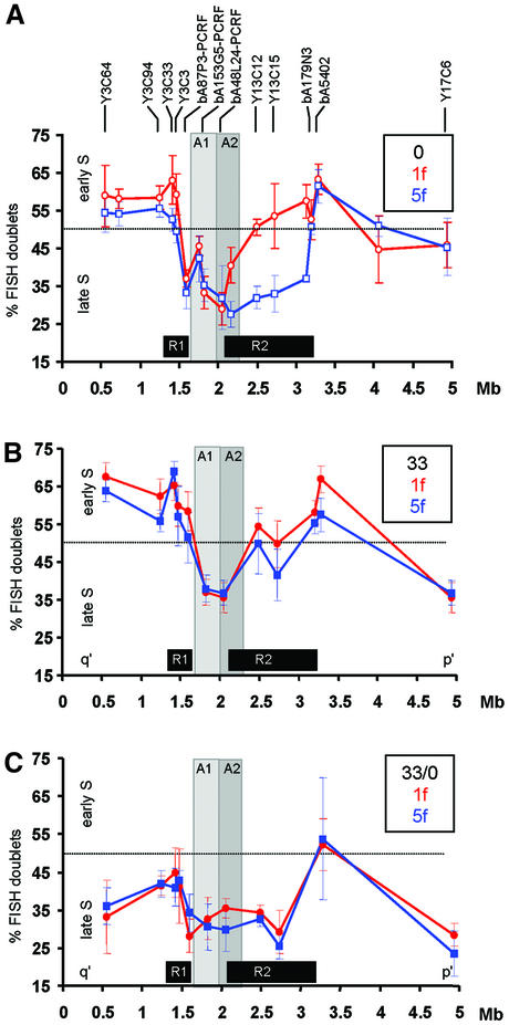
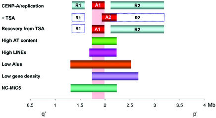
We show that Trichostatin A (TSA)-induced partial histone hyperacetylation causes a unidirectional shift in the position of a previously defined binding domain for the centromere-specific histone H3 homologue CENP-A at a human neocentromere. The shift of approximately 320 kb is fully reversible when TSA is removed, but is accompanied by an apparent reduction in the density of CENP-A per unit length of genomic DNA at the neocentromere. TSA treatment also instigates a reversible abolition of a previously defined major domain of differentially delayed replication timing that was originally established at the neocentromeric site. None of these changes has any measurable deleterious effects on mitosis or neocentromere function. The data suggest pliability of centromeric chromatin in response to epigenetic triggers, and the non-essential nature of the regions of delayed replication for centromere function. Reversibility of the CENP-A-binding position and the predominant region of delayed replication timing following removal of TSA suggest strong memory at the original site of neocentromeric chromatin formation.


Figures

None
Fig. 1. Effects of TSA concentration on cell growth and chromosome segregation. (A) Doubling time was determined by maintaining cells in exponential phase (with splitting every 2 days) for a total of 6 days. Results were averaged from six separate experiments. (B) Viability was determined by Trypan Blue staining from a sample of cells during each of the culture splits in (A). (C) The level of missegregation was calculated by scoring 50 cells for the number of anaphase II/telophase cells exhibiting lagging or bridging chromosomes. Results averaged from four separate experiments were divided by those of untreated cells to give relative levels of missegregation. (D) The level of micronuclei was calculated by scoring 1000 cells in four separate experiments and dividing the value by that of the untreated cells.
None
Fig. 2. Effects of TSA concentration on cellular levels of acetylated histones H4 (AcH4) and/or H3 (AcH3). (A and B) Western blotting. (A) 5f cells were cultured without TSA or with 33, 150 or 300 nM TSA for 17 h. In some experiments (33/0), cells were grown in the presence of 33 nM TSA for 17 h, washed, and grown in TSA-free medium for a further 3 days. Untreated controls (0) were grown for the same length of time. Total cell lysates from equivalent numbers of cells were run on denaturing SDS–PAGE and blotted with antibodies against acetylated histone H4 (AcH4), histone H3 (AcH3) or CENP-A. In all cases, equal gel loadings were confirmed using an anti-β-tubulin antibody. (B) Quantitation of band intensity in (A) for 0, 33, 150, 300 and 33/0 nM TSA for acetylated histones. (C and D) Immunofluorescence. (C) Metaphase chromosomes (DAPI stained in upper panels) from unfixed and cytospun 5f cells cultured as in (A) were stained with an antibody against acetylated histone H4 (lower panels). Bar, 10 µm. (D) Quantitation of fluorescence intensities in (C) at the centromeres and non-centromeric regions following TSA treatment. Signal intensities in (B) and (D) were given arbitrary units after correcting for background signals on the gels or slides.
None
Fig. 3. Effects of TSA on CENP-A distribution at the mardel(10) neocentromere. (A) No TSA treatment (0; data taken from Lo et al., 2001a). (B) Treatment with 33 nM TSA for 17 h (33). (C) Treatment with 33 nM TSA for 17 h followed by 3 days without TSA (33/0). Data were collected using a previously described CIA analysis procedure (Lo et al., 2001a,b). Percent differences between the normalized values of 5f [mardel(10)] and 1f (normal chromosome 10) are plotted against the midpoint position of each BAC within the 5 Mb 10q25 BAC contig described previously (Lo et al., 2001a). Each data point is the mean of two to five independent experiments. All data points are shown with error bars (±SE). The grey box shows the CENP-A binding region defined in (A) (Lo et al., 2001a). Experiments for the untreated cells reported previously (Lo et al., 2001a,b) and the TSA-treated cells presented here were performed, and the two data sets collected and analysed, simultaneously. The designations for several pertinent BACs are shown.
None
Fig. 4. Replication timing at the 10q25 neocentromere region. (A) No TSA treatment (0; data taken from Lo et al., 2001a). (B) Cells treated for 17 h with 33 nM TSA (33). (C) Cells treated for 17 h with 33 nM TSA followed by culture without TSA for 3 days (33/0). Average percentages of S phase FISH signal doublets (inversely proportional to replication time) for each probe are plotted against the midpoint position of each probe (shown above the graph) within the 5 Mb 10q25 contig for 1f (normal chromosome 10; red circles) and 5f [mardel(10); blue squares]. Results from untreated cells (0; Lo et al., 2001a) are shown with open symbols; results from TSA-treated cells obtained in the present study are shown as filled symbols. The dotted horizontal line at 50% FISH doublets represents the approximate mid-S phase time-point. Minimal CENP-A-binding domains before and after TSA-treatment are designated A1 and A2 and shown as light and darker grey areas, respectively. R1 and R2 boxes denote the regions of differentially delayed replication on mardel(10) compared with normal chromosome 10 in the absence of TSA (Lo et al., 2001a). Results are the averages ±1 SD of four separate experiments. Experiments for the untreated cells reported previously (Lo et al., 2001a) and the TSA-treated cells presented here were performed, and the two data sets collected and analysed, simultaneously.
None
Fig. 5. Summary of the distribution of different domains at the 10q25 neocentromere region. A1 designates the position of the minimal CENP-A-binding domain before TSA (Lo et al., 2001a) or following 3 days of recovery from 33 nM TSA treatment, whereas A2 denotes the position of the minimal CENP-A-binding domain after 17 h of 33 nM TSA treatment. Solid R1 and R2 boxes denote the regions of delayed replication on mardel(10) compared with normal chromosome 10 in the absence of TSA (Lo et al., 2001a). Open R1 and R2 boxes denote the same regions whose delayed replication characteristics are abolished following treatment with TSA, noting that the major domain R2 (but not R1) is reversible after removal of TSA. Regions of high AT (>62%) and LINE interspersed repeat contents and regions of depressed Alu repeats and predicted gene contents are shown (Lo et al., 2001a). NC-MiC5 represents a 0.65 Mb stable minichromosome previously constructed in our laboratory (Saffery et al., 2001).

References

    1. Aagaard L., Schmid,M., Warburton,P. and Jenuwein,T. (2000) Mitotic phosphorylation of SUV39H1, a novel component of active centromeres, coincides with transient accumulation at mammalian centromeres. J. Cell Sci., 113, 817–829. - PubMed
    1. Ahmad K. and Henikoff,S. (2001) Centromeres are specialized replication domains in heterochromatin. J. Cell Biol., 153, 101–110. - PMC - PubMed
    1. Barry A.E., Howman,E.V., Cancilla,M.R., Saffery,R. and Choo,K.H.A. (1999) Sequence analysis of an 80 kb human neocentromere. Hum. Mol. Genet., 8, 217–227. - PubMed
    1. Barry A.E., Bateman,M., Howman,E.V., Cancilla,M.R., Tainton,K.M., Irvine,D.V., Saffery,R. and Choo,K.H.A. (2000) The 10q25 neocentromere and its inactive progenitor have identical primary nucleotide sequence: further evidence for epigenetic modification. Genome Res., 10, 832–838. - PMC - PubMed
    1. Bickmore W.A. and Carothers,A.D. (1995) Factors affecting the timing and imprinting of replication on a mammalian chromosome. J. Cell Sci., 108, 2801–2809. - PubMed

Publication types